|Poll|November 14, 2013
Image IQ: 47-year-old with Irregular Mass in Dense Breast Tissue
Author(s)Stamatia Destounis, MD, FACR
Advertisement
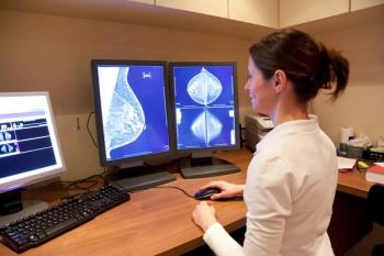
A 47-year-old woman presents for screening mammography and requests a screening breast ultrasound. On mammography, no suspicious masses or areas of calcifications are identified. The breast tissue is extremely dense.
On ultrasound, there is a newly identified irregular hypoechoic mass in the left 2:00 area.
(Click on images to enlarge.)
What is your diagnosis?
Newsletter
Stay at the forefront of radiology with the Diagnostic Imaging newsletter, delivering the latest news, clinical insights, and imaging advancements for today’s radiologists.
Advertisement
Latest CME
Advertisement
Advertisement
Trending on Diagnostic Imaging
1
Leading Breast Radiologists Discuss the Recent Lancet Study on AI and Interval Breast Cancer
2
Is AI Better Than Neuroradiologists at Evaluating Aneurysm Growth on CTA and MRA Scans?
3
FDA Clears AI-Powered Triage Platform for Digital Breast Tomosynthesis
4
Study Shows Photon-Counting CT Reduces Radiation Exposure by 66 Percent for Patients with Lung Cancer
5


