|Articles|January 6, 2006
Intraprocedural CT helps eliminate recurrent kidney tumors
Intraprocedural contrast-enhanced CT helped reduce the rate of incompletely treated renal cell carcinoma lesions by approximately 64%, according to a study presented during the Interventional Oncology Symposium at the 2005 RSNA meeting.
Advertisement
Intraprocedural contrast-enhanced CT helped reduce the rate of incompletely treated renal cell carcinoma lesions by approximately 64%, according to a study presented during the Interventional Oncology Symposium at the 2005 RSNA meeting.
The retrospective study of 90 tumors in 82 patients with stage 1 and 2 renal cell carcinoma divided patients into two groups. Patients with creatinine levels of 2 or less underwent a contrast CT study after the tumor was presumed to be fully ablated. Patients with a creatinine level higher than 2 were not scanned, and the ablation endpoint was determined by the estimated ablation zone.
The intraprocedural scans found residual tumors in 10% of patients, said lead author Dr. Kyle A. Krehbiel of Wake Forest University. The team used contrast CT to target the residual tumor and immediately reablated the area.
After an average follow-up of 10 months, the team discovered recurrent disease in 5.8% of patients who had undergone intraprocedural contrast CT. By comparison, 19% of the patients who had not undergone intraprocedural contrast CT had recurrent disease.
On follow-up, none of the residual tumors that had required additional ablation had recurred. However, 6.4% of tumors that did not enhance with contrast CT showed recurrent disease on follow-up imaging, indicating that the scans may miss microscopic viable tumor cells.
The size of the lesion seemed to influence the rate of recurrence. The average size of lesions in either group that showed residual or recurrent tumor was 4.3 cm, while lesions that did not show residual or recurrent tumor averaged 2.5 cm.
For more online information, refer to Diagnostic Imaging's
Advertisement
Related Content
Advertisement


Advertisement
Advertisement
Trending on Diagnostic Imaging
1
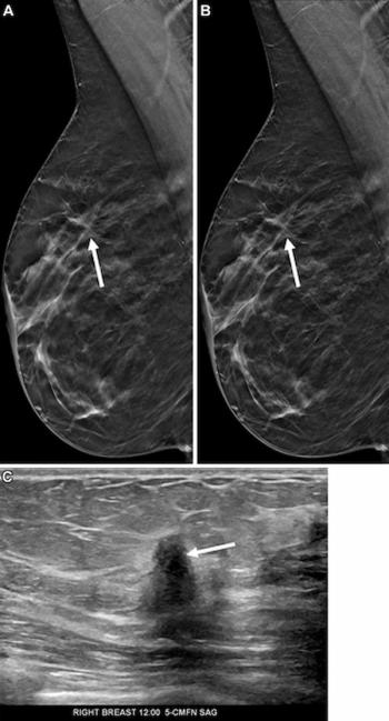
Mammography Study Shows Impact of AI-Powered Slab Reconstruction with DBT
2
GE HealthCare Unveils 4D Dynamic PET Imaging Software at SNMMI Conference
3
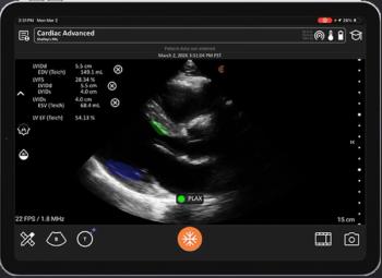
FDA Clears AI Ultrasound Software for Automated LVEF Calculation
4
Can AI Assessment of CT Attenuation Correction Mapping Be a Viable Alternative for CAC Risk Stratification?
5