Mammography Study Shows Link Between Enlarged Axillary Lymph Nodes and Higher Risks for Diabetes and Cardiovascular Disease
The presence of fat-enlarged axillary lymph nodes on mammography screening exams is associated with a fourfold higher risk of type 2 diabetes and a 2.6-fold higher risk for cardiovascular disease, according to new research presented at the ARRS Annual Meeting.
Emerging research suggests the detection of fat-enlarged axillary lymph nodes on screening mammography exams may be associated with significantly higher risks of type 2 diabetes (T2DM), cardiovascular disease (CVD), and hypertension (HTN).
In a retrospective study, which garnered Summa Cum Laude honors at the 2024 American Roentgen Ray Society (ARRS) Annual Meeting in Boston, researchers reviewed bilateral mediolateral oblique views from mammography exams for 1,216 women who had cardiovascular risk factors within one year of the mammogram but no known coronary artery disease (CAD) at the time of the mammography screening. The study authors measured the largest visible axillary lymph node and also determined if the patients had any major adverse cardiovascular events (MACEs) within 10 years after the index mammogram.
The researchers found that 19.1 percent of the cohort had fat-enlarged axillary lymph nodes (greater than 20 mm in length) due to an expanded fatty hilium. For these patients, the study authors noted a fourfold higher risk for T2DM, a 2.6-fold higher risk of CVD, and a 2.5-fold higher risk of HTN.


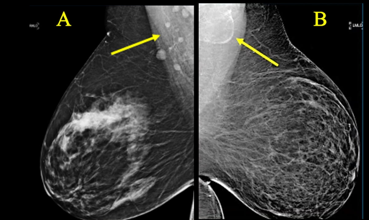
Here one can see a comparison between mammography findings for two women with similar body mass index (BMI). Note the normal axillary lymph nodes in a 63-year-old woman (A) and a fat-enlarged axillary lymph node in a 52-year-old woman (B). New research revealed a fourfold higher risk of type 2 diabetes and a 2.6-fold higher risk for cardiovascular disease in women presenting with fat-enlarged axillary lymph nodes on mammography exams. (Images courtesy of the American Roentgen Ray Society (ARRS).
“Fat-enlarged axillary lymph nodes visualized on screening mammography may increase the ability to identify women who would benefit from CVD risk reduction strategies and more intensive risk assessment with coronary artery CT,” suggested lead study author Jessica Rubino, M.D., who is affiliated with the Dartmouth Hitchcock Medical Center in Lebanon, N.H., and colleagues.
The researchers noted that axillary lymph nodes were visible on 907 of the 1216 mammography exams reviewed in the study (74.6 percent). Emphasizing the wide eligibility for screening mammography for over 75 percent of women in the United States, the study authors emphasized that obtaining cardiovascular risk data from mammograms could have a significant impact in reducing mortality from CVD, the leading cause of death in women.
“If validated in larger studies, incorporating fat-enlarged nodes into CVD risk models has the potential to improve CVD risk stratification without additional cost or additional testing,” posited Rubino and colleagues.
Reference
1. Rubino J, Austin-Strohbehn J, Wang Q, et al. Fat-enlarged axillary lymph nodes on screening mammograms predict cardiometabolic disease and cardiovascular disease risk. Presented at the 2024 American Roentgen Ray Society (ARRS) Annual Meeting in Boston, May 5-9, Boston. Available at: https://apps.arrs.org/MeetingPortal24/ . Accessed May 5, 2024.
Newsletter
Stay at the forefront of radiology with the Diagnostic Imaging newsletter, delivering the latest news, clinical insights, and imaging advancements for today’s radiologists.

 and Parkinson-Plus Syndrome")


Mammography Study: AI Facilitates Greater Accuracy and Longer Fixation Time on Suspicious Areas
July 8th 2025While noting no differences in sensitivity, specificity or reading time with adjunctive AI for mammography screening, the authors of a new study noted a 4 percent higher AUC and increased fixation time on lesion regions.


SNMMI: Can 18F-Fluciclovine PET/CT Bolster Detection of PCa Recurrence in the Prostate Bed?
June 24th 2025In an ongoing prospective study of patients with biochemical recurrence of PCa and an initial negative PSMA PET/CT, preliminary findings revealed positive 18F-fluciclovine PET/CT scans in over 54 percent of the cohort, according to a recent poster presentation at the SNMMI conference.

Could an Emerging PET Tracer be a Game Changer for Detecting Hepatocellular Carcinoma?
June 23rd 2025In addition to over 90 percent sensitivity in detecting hepatocellular carcinoma (HCC), the glypican-3 (GPC3) targeted PET tracer 68Ga-aGPC3-scFv appeared to be advantageous in identifying HCC tumors smaller than one centimeter, according to pilot study findings presented at the SNMMI conference.

