While adjunctive AI and mammography did offer a significantly higher specificity rate in comparison to the use of supplemental breast ultrasound (95.3 percent vs. 77.6 percent), the researchers found the combination of ultrasound and mammography was more adept at detecting smaller, early node-negative breast cancer in women with dense breasts.
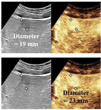
Out of the 12 cases of breast cancer missed by adjunctive AI and detected with supplemental ultrasound, half of the cases involved ductal carcinoma in situ (DCIS) and five cases were stage 1 invasive cancer. The researchers also noted the average size of the additional cancer lesions detected with supplemental ultrasound was 0.3 cm2 in comparison to 1.0 cm2 for mammography/AI detected breast cancer lesions.
“ … From an early cancer detection standpoint, offering supplemental breast (ultrasound) for women with dense breasts, even after a negative assessment according to AI, could be a method to reduce the number of interval cancers,” posited Ha and colleagues.
Three Key Takeaways
1. Higher sensitivity with ultrasound. The combination of supplemental breast ultrasound and mammography significantly improves the sensitivity rate for breast cancer detection (97 percent) compared to the combination of mammography and adjunctive AI (60.6 percent) in women with dense breasts.
2. Detection of additional cancers. Supplemental ultrasound helped detect additional cancers, including smaller and early-stage breast cancers that mammography with AI missed. This included 12 additional cancers, with half being ductal carcinoma in situ (DCIS) and five cases of stage 1 invasive cancers.
3. Higher abnormal interpretation rate (AIR) and cancer detection rate (CDR). The use of supplemental ultrasound had a higher AIR percentage (22.9 percent vs. 5 percent) and CDR (5.6 vs. 3.5 per 1,000 exams) compared to adjunctive AI, indicating improved cancer detection capabilities, despite adjunctive AI offering higher specificity (95.3 percent vs. 77.6 percent).
In an accompanying editorial, Gary J. Whitman, M.D., and Stamatia V. Destounis, M.D., noted that AI can play a key role in breast cancer screening. However, based upon the recent literature and the current study findings, they emphasized caution with the use of AI in women with dense breasts and further research.
“These findings suggest that AI may not improve the performance of mammography in women with dense breasts, and additional larger well-designed studies are needed to verify these findings,” maintained Dr. Whitman, a professor of breast imaging and breast radiation oncology at the University of Texas MD Anderson Cancer Center, and Dr. Destounis, a managing partner at Elizabeth Wende Breast Care in Rochester, N.Y., and the chair of the American College of Radiology (ACR) Breast Commission.
(Editor’s note: For related content, see “What a New Study Reveals About Whole-Breast Ultrasound Tomography, Mammography and Dense Breasts,” “Leading Breast Radiologists Discuss the USPSTF Breast Cancer Screening Recommendations” and “Study of Mammography AI Software Notes 50 Percent Higher Likelihood of False-Positive Results for Black Women.”)
Beyond the inherent limitations of a single-center retrospective study, the authors acknowledged that 90 percent of the cohort had low or average risk for breast cancer. They also pointed out that reviewing radiologists did not have access to clinical information and prior imaging exams. Given that their results involved one AI system, the researchers cautioned against broad application of the study findings to other AI systems.