|Articles|October 29, 2003
Molecular medicine drives demand for animal scanners
Preclinical studies help predict imaging needsEquipment manufacturers are taking an increasing interest in the market for dedicated animal scanners. The development of molecular medicine, driven by advances in proteomics and
Advertisement
Preclinical studies help predict imaging needs
Equipment manufacturers are taking an increasing interest in the market for dedicated animal scanners. The development of molecular medicine, driven by advances in proteomics and genomics, is bringing the traditionally separate areas of clinical and preclinical imaging closer than ever before.
Two recent international meetings highlighted the growing significance of animal imaging data to proposed "smart" drugs and novel gene therapies. Presenters at the Society of Noninvasive Imaging in Drug Development meeting, held in Madrid in September, noted that genomic research has identified countless targets in the human body that drug treatments and gene therapy could address, while advanced combinatorial chemistry techniques are generating millions of complex molecular compounds with unknown functional properties. Matching the right pharmaceutical with an appropriate target is not only enormously difficult and time-consuming, it is also extremely costly if decisions turn out to be incorrect. Animal testing will play a pivotal role, and vendors have decided dedicated animal scanners are worth pursuing.
Molecular imaging studies on rats and genetically engineered mice have the potential to identify likely winners and losers at an early stage of the lengthy drug trial process. The noninvasive techniques also let researchers assess the effects of repeated dosing regimens. Consequently, this drug development process is opening up a ready market for small-scale imaging equipment at industrial and academic research centers.
"The sheer volume of molecules that can be synthesized and must be tested in animal models will grow the market for animal scanners," said Jonathan Frey, manager of PET marketing for Siemens Medical Solutions. "The ability to perform serial studies, through both diagnosis and therapy, will speed the drug development process and reduce the cost of trials."
The imaging industry's top medical imaging executives, gathered in Paris for August's annual meeting of the International Society for Strategic Studies in Radiology (ISSSR), agreed that diversifying into animal imaging could reap rewards.
"There will be more rats scanned (in molecular imaging) than people in the next 10 years," said Joseph M. Hogan, president and CEO of GE Medical Systems.
Executives present at the ISSSR meeting acknowledged that support for in vivo preclinical research will directly feed into future clinical imaging applications, both in diagnostics and therapy-monitoring.
"We are convinced that molecular medicine will make a significant contribution toward the development of healthcare," said Dr. Erich R. Reinhardt, president of Siemens Medical Solutions. "But in order to develop the human molecular imaging applications, you have to utilize the animal molecular imaging techniques."
Michael Licata, general manager of PET preclinical imaging systems for Philips Medical Systems, is equally confident that medical imaging companies should take advantage of synergies between the preclinical and clinical worlds.
"There is a paradigm shift happening in imaging," Licata said. "We're working together with researchers and universities worldwide in their molecular imaging developments. It's like having a crystal ball to see what the clinical imaging requirements will be in the future."
All three medical imaging giants have moved aggressivley into the animal imaging market over the past 12 months. In November 2002, GE acquired Ontario-based Enhanced Vision Systems, a pioneer in micro-CT technology. GE's micro-CT product portfolio includes the eXplore RS, which is designed to image small laboratory animals as well as in vitro biomedical specimens. The company has also signed a strategic alliance with ART Advanced Research Technologies to develop new optical molecular imaging applications that will initially be applied in animal studies.
Philips went public with its own small-bore PET scanner, the Mosaic, at the Society of Nuclear Medicine's 50th annual meeting in June. Philips also chose the SNM meeting to announce an exclusive worldwide distribution agreement with ImTek of Knoxville regarding its MicroCAT II in vivo CT scanner for use in animal research.
One month later, Siemens signed an agreement with Gamma Medica, a California nuclear imaging company. The July deal secured Siemens exclusive U.S. marketing rights to Gamma Medica's X-SPECT, which can be configured as a single- or dual-head animal imaging system. Siemens is continuing to market its own M.CAM system, a large field-of-view camera adapted for small animal planar imaging. The company also distributes MicroPET products developed by Knoxville-based Concorde Microsystems for large and small animal imaging.
It is no coincidence that each of the major medical imaging companies is investing in systems that generate functional and anatomical information, either together or separately. This approach mirrors the trend toward combining data in human molecular imaging studies. The morphing of high-resolution 2D and 3D body scans onto color maps of metabolic activity lets researchers pinpoint the site and level of physiological response accurately, regardless of whether the subject is a mouse or a human.
X-SPECT, for example, can be coupled with data obtained using a miniature CT, generating coregistered CT and SPECT images. No combined dedicated animal PET/CT yet exists, though Philips has taken steps to optimize compatibility between the Mosaic small-bore PET and ImTek's MicroCAT system.
"Our pallet animal-holding device can be interchanged between the two systems," Licata said. "We're also checking that all the DICOM communication between the systems works."
Philips has designed its small-bore PET scanner to be as similar to its clinical PET systems as possible, using the same software platform. This will enable meaningful comparisons between preclinical and clinical data, Licata said. Researchers will also be able to concentrate on imaging studies, rather than scanner protocols.
"Animal imaging is not new, but today we have a different type of researcher using the imaging equipment. It's no longer the imaging expert or imaging scientist, it's a chemist or biologist," he said. "These folks want to treat the animal scanner like a tool on their workbench, so we're working to have our systems do exactly that. It's a research tool with clinical capabilities, reliability, and robustness."
Philips' formal entry into the preclinical imaging market in June was preceded by two years of intensive R&D and market research, Licata said. He is optimistic that the company's investment will be returned as global market potential is realized. Mosaic small-bore PET systems are now up and running at two trial sites: the University of Pennsylvania and the Peter MacCallum Cancer Center in Melbourne, Australia. Philips began taking orders for the product at the SNM meeting and aims to ship its first commercial rodent PET scanners before the end of 2003.
"I think that in 2004, and certainly by 2005, we'll start to see the animal imaging business itself being profitable," Licata said.
The company is also investigating further opportunities in animal imaging in other modalities. Preclinical imaging is a multimodality enterprise. SPECT, optical imaging, MRI, and even ultrasound will all have a role to play, Licata said.
"All the imaging modalities are going to be important to the animal researcher because they each provide a different piece of the puzzle, or a different form of the information they need for their research," he said.
Advertisement
Related Content
Advertisement


Advertisement
Advertisement
Trending on Diagnostic Imaging
1
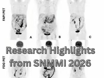
Molecular Imaging in Focus: Key Takeaways from the 2026 SNMMI Conference with Michael Hofman, MBBS, FRACP
2
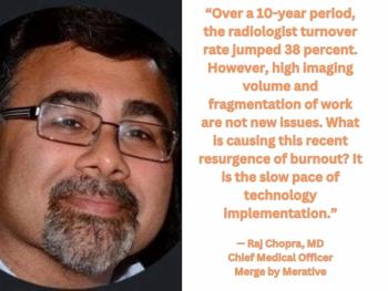
Five Strategies to Facilitate Technology Implementation and Alleviate Radiologist Turnover
3
FDA Approves Low-Dose MRI Contrast Agent Gadoquatrane
4
Can Breast Ultrasound AI Offer Comparable Sensitivity to Radiologists in Pregnant and Lactating Women?
5