MRI vendors pursue real-time imaging with interactive scanning protocols
New techniques could spur interventional usesControl has taken its place alongside speed as a major driver of new MR technologies. This control is taking the form of interactivity: the ability to control data acquisition moment to moment, which is
New techniques could spur interventional uses
Control has taken its place alongside speed as a major driver of new MR technologies. This control is taking the form of interactivity: the ability to control data acquisition moment to moment, which is a major step toward making MR a real-time imaging modality.
MR vendors demonstrated their progress toward interactive imaging at last months RSNA meeting. On display were scanners, peripherals, including phased-array coils, and software packages designed to make the most productive use of real-time imaging. The resultant images displayed in booths on the RSNA exhibit floor were remarkable, not only for their clarity and resolution, but also for their ability to visualize parts of the body beyond the reach of most installed MR systems.
Both GE and Siemens are promoting the use of interactivity as the way to realize the true potential of real-time imaging. Gains in productivity and expanded interventional capability could be the most immediate results.
Until now, faster imaging has involved repeating the same slice over and over to watch events happening within that slice, said John Pavlidis, manager of the Siemens MR division in Iselin, NJ. Interactive capability gives us control over the slice position.
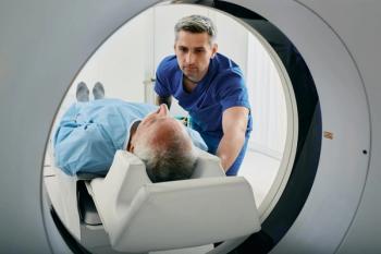
With interactive tools, operators can increase or decrease contrast to better visualize anomalies or change the scan plane being viewed on the fly. Diagnostically, such changes may be used to focus on damage from a suspected stroke or heart attack, for example. When enough information is in hand to make the diagnosis, the exam can be stopped. As an interventional tool, interactivity promises to allow unprecedented visualization of interventional probes and surrounding tissue.
To harness interactivity, MRI engineers have developed special tools that allow operators to navigate through the body. One approach is to look at different slices in real time as part of the diagnostic process, tailoring protocols to gather the most relevant information.
In this way, were using the MR system like an ultrasound scanner, Pavlidis said.
The accumulated data can also be assembled off-line into volumes, from which specific slices can be extracted for more detailed examination. Or, slices can be displayed as an integrated whole. The result can be a dynamic model of the heart completing a cycle. This 3-D model can then be taken apart for qualitative assessment or quantitative measurement.
One such cardiac model created with GE technology clearly depicts abnormal wall motion. With the administration of a contrast agent, the area of the heart not getting enough blood shows up as a dark spot.
GE optimized its flagship MR scanner for this kind of high-speed imaging. The result was Signa CV/i (cardiovascular interactive). Interactivity is a means for delivering on the promise of high-speed MR imaging, according to Eugene Saragnese, general manager of global MR for the Milwaukee company.
The ability to make scan-plane adjustments and image processing on the fly are key elements in a number of new applications. Its important in cardiac imaging, where you need to localize planes, or when doing peripheral vascular run-offs with a single bolus, from the renal (arteries) down to the feet, Saragnese said.
GE is developing an eight-coil peripheral vascular array specifically for such run-off studies. Interactivity provides operators with the ability to chase the contrast bolus as it moves through the patient, and users can switch from one phased-array coil to the next. The array, which is pending Food and Drug Administration clearance, could be shipping before the end of the first quarter.
While interactivity has its greatest impact at high-field, opportunities exist at mid-field as well. GE is implementing versions of the hardware and software required to support interactivity for use on the Signa Profile/i. Later this year, the mid-field system will be equipped with high-performance phased-array coils and new gradients to allow high-speed imaging. The software for controlling the data is already in place.
GE executives expect the system to be used primarily for diagnostic applications, but the company is also developing peripherals that will support its use for interventional procedures. Among the additions are an in-room monitor and controls for using the interactive capabilities during intervention.
There is a potential downside to interactivity. Increasing the involvement of the operator raises the chance of slowing down procedures. The solution is to automate this interactivity, just as current interfaces have been developed to be more user-friendly. For example, Picker International of Cleveland is developing technology that will automatically track biopsy needles or probes as they move through tissue.
If the scanner knows the location of the biopsy needle, you should be able to tell it to continue scanning in that plane, or maybe 2 cm in front, said Linda Eastwood, marketing manager of Pickers MR division. Why should a technologist have to figure out which scan plane to be in?
While interactivity is appealing, its true impact has yet to be understood. In recent years, engineers have focused on building MR scanners that require less technologist training. Interactivity, therefore, might be initially adopted by facilities with the best-trained staff. There is, however, little doubt that this new capability will spur innovative clinical approaches for MR, and, as the clinical utility of these applications is established, new user-friendly techniques will emerge.
Our customers are applications-based, Saragnese said. Interactivity is a key enabler and applications is what it delivers.
Newsletter
Stay at the forefront of radiology with the Diagnostic Imaging newsletter, delivering the latest news, clinical insights, and imaging advancements for today’s radiologists.


