
|Articles|February 17, 2020
PET Scans Identify Brain Inflammation in Veterans with Gulf War Illness
Author(s)Whitney J. Palmer
Neuroinflammation discovered in regions of the brain that control higher-order functions.
Advertisement
Veterans diagnosed with Gulf War Illness (GWI) have significantly higher elevations of brain inflammation than other individuals – a finding from a new study that could support treating neuroinflammation as a potential therapeutic target.
These soldiers experience neuroinflammation in areas of the brain that are responsible for executive functions, including memory, concentration, and reasoning. This symptom was similar to what is seen in patients with fibromyalgia, according to researchers from Massachusetts General Hospital’s Athinoula A. Martinos Center for Biomedical Imaging led by Marco Loggia, Ph.D., associate professor of radiology at Harvard Medical School.
The study, published in
GWI affects approximately 30 percent of soldiers who served during the 1991 Gulf War. It prompts fatigue, chronic pain, and cognitive problems, such as memory loss, but the root cause remains unknown. Some investigator have hypothesized the condition could result from exposure to nerve gas and the medication that counteracts the neurotoxin, pesticide exposure, sleep deprivation, stress associated with extreme temperature changes, and physical exertion during deployment.
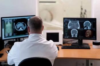
To determine whether veterans with GWI experience neuroinflammation like fibromyalgia patients, Loggia’s team used PET imaging to scan the brains of 23 veterans, 15 who had GWI and 25 healthy civilians. The scans used the radioligand [11C]PBR28 to track and measure the levels of translocator protein, a molecule that increases in amount in the glial cells that surround and insulate neurons during the neuroinflammatory response. The areas that had the most inflammation were the precuneus, prefrontal cortex, and primary motor and somato-sensory areas.
The imaging revealed veterans with GWI had more extensive neuroinflammation than their healthy counterparts. Further research with a larger study population is needed to validate the results and determine whether glial modulation could be an effective therapy for these soldiers.
These findings also add to the existing body of knowledge on the implications of neuroinflammation from Loggia’s lab. Prior research indicates neuroinflammation also plays a role in many other conditions, including chronic pain, depression, anxiety, and autism. In addition, it could be integral to amyotrophic lateral sclerosis (ALS), multiple sclerosis, Huntington’s disease and migraine.
The hope, Loggia said, is that the findings with veterans with GWI “could help motivate a more aggressive evaluation of neuroinflammation as a potential therapeutic target.”
Advertisement
Related Content
Advertisement


Advertisement
Advertisement
Trending on Diagnostic Imaging
1
Can Breast Ultrasound AI Offer Comparable Sensitivity to Radiologists in Pregnant and Lactating Women?
2
Molecular Imaging in Focus: Heather Jacene, MD, Charts Priorities as the New President of SNMMI
3
FDA Approves Low-Dose MRI Contrast Agent Gadoquatrane
4
Molecular Imaging in Focus: Key Takeaways from the 2026 SNMMI Conference with Michael Hofman, MBBS, FRACP
5