
|Articles|May 25, 2007
Philips explores colorectal screening with fluoro-based MRI
Philips Medical Systems is assessing the use of naturally occurring, nonradioactive fluorine-19 as part of an MR contrast agent that will root out the early signs of colorectal cancer.
Advertisement
Philips Medical Systems is assessing the use of naturally occurring, nonradioactive fluorine-19 as part of an MR contrast agent that will root out the early signs of colorectal cancer.
The company unveiled in Berlin a research program to identify biomarkers specific to this type of cancer and attach them to microcapsules filled with stable and nontoxic forms of fluorine. The research into biomarkers and the ligands for attaching them to the fluorine is being done in collaboration with academic partners. Development of the nanoparticles and their visualization is being done in-house at the company's Life Sciences facilities in Eindhoven, the Netherlands, using a 3T Achieva scanner to conduct the preclinical studies.
The company chose the scanner to replicate the scanning environment in which the agent, when completely developed, will be examined.
"If you develop an agent at a higher field and then go to a lower one in a clinical setting, the contrast medium may have very different behavior and may not work as expected," said Rolf Lamerichs, Ph.D., senior scientist in the biomolecular engineering division of Philips Research. "With the Achieva, we have the sequences available and performance is robust."
Once developed, the contrast agent - composed of a fluorine microcapsule and ligand binding the capsule to a biomarker such as an antibody specific to colorectal lesions - would be administered orally. This agent would wash over the intestinal tract, attaching selectively to precancerous adenomas and cancers. Preliminary research indicates that two highly specific contrast agents might be possible, one for precancerous and the other for cancerous lesions.
Excess nanoparticles would then be flushed out, leaving fluorine hot spots, to be identified using a technique developed by Philips called fluorine ultrafast turbo spectroscopic imaging. F-uTSI uses spectral information to home in on the presence of the F-19 nanoparticles, while correcting for artifacts that commonly occur due to the imaging of multiple resonance frequencies associated with fluorine. The corrected data would then be reconstructed into images of the contrast agent and overlaid onto anatomic images of the intestinal tract, which would provide the context for localizing lesions.
"The plan is to replace unfriendly colonoscopy with a better-tolerated MRI procedure," Lamerichs said.
Much remains to be done, particularly in the development of the biomarker. Philips has covered a lot of ground already, however, coming up with ways to package and visualize the fluorine.
"When these agents come, we will be able to image them in the proper and correct way," he said.
Advertisement
Related Content
Advertisement


Advertisement
Advertisement
Trending on Diagnostic Imaging
1
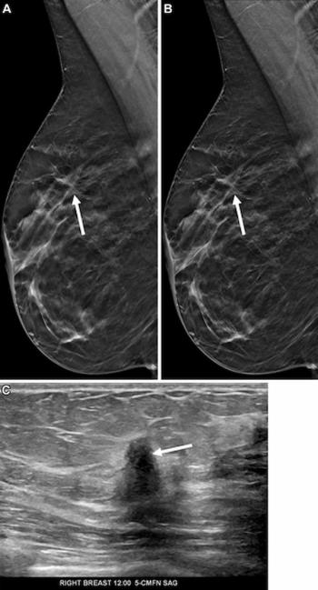
Mammography Study Shows Impact of AI-Powered Slab Reconstruction with DBT
2
GE HealthCare Unveils 4D Dynamic PET Imaging Software at SNMMI Conference
3
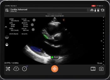
FDA Clears AI Ultrasound Software for Automated LVEF Calculation
4
Can AI Assessment of CT Attenuation Correction Mapping Be a Viable Alternative for CAC Risk Stratification?
5