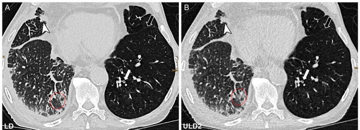
A new study suggests the use of ultralow-dose photon counting computed tomography (ULD PCT) may offer sufficient diagnostic quality and adequate anatomical visualization of lung deformities in patients who have undergone a lung transplant.
For the prospective study, recently reported in Radiology, researchers compared low-dose computed tomography (LDCT) at 1.41 mSv and two ULD PCT protocols at .08 mSV (ULD1) and .17 mSV (ULD2) for obtaining computed tomography (CT) images in 82 patients (median age of 64) who had lung transplants.
The study authors found that reviewing radiologists noted sufficient image quality (Likert score > 3) for diagnosis in 97.5 percent of patients who had the ULD1 protocol (39/40 patients) and 97.6 percent of those who had the ULD2 protocol (40/41 patients). Adequate anatomical visualization (Likert score > 3) was achieved for 80.5 percent of patients who had the ULD1 protocol (33/41) and 82.9 percent of patients receiving the ULD2 protocol (34/41), according to the researchers.
“ … This study provides evidence that ultralow-dose (ULD) photon-counting CT (PCT) is a promising tool for repeat follow-up chest CT in recipients of lung transplant at only one-tenth the radiation dose of a standard low-dose CT examination, with the potential for a reduction in cumulative life-time radiation exposure,” wrote lead study author Ruxandra-Iulia Milos, M.D., who is affiliated with the Departments of Biomedical Imaging and Image-Guided Therapy at the Medical University of Vienna in Vienna, Austria, and colleagues.
The researchers also noted little difference between the ULD1 and ULD2 protocols with respect to image noise (74.37 Hounsfield units (HU) vs. 74.28 HU) and the signal-to-noise ratio (SNR) (18.73 vs. 18.89).
Three Key Takeaways
1. Radiation dose reduction. Ultralow-dose photon-counting CT (ULD PCT) significantly reduces radiation exposure, delivering diagnostic images at just one-tenth of the dose of standard low-dose CT, which is important for repeat follow-up imaging in lung transplant patients.
2. Diagnostic accuracy. Both the ULD1 (0.08 mSv) and ULD2 (0.17 mSv) protocols provided sufficient image quality for diagnosis in over 97 percent of cases, with comparable sensitivity and specificity for detecting pulmonary pathologies.
3. Similar image quality with lower radiation dose. Despite the lower radiation doses, image noise and signal-to-noise ratios (SNRs) were almost identical between ULD1 and ULD2 protocols, making them viable alternatives to traditional low-dose CT for anatomical visualization and pathology identification.
“Despite substantial dose reduction, image noise and the signal-to-noise ratios were similar between the two ULD protocols, and the ULD protocols were accurate for the identification of pulmonary pathologies with sensitivity and specificity comparable to previously published data on ULD CT,” maintained Milos and colleagues.
For the LDCT images, there was substantial interobserver agreement on consolidations, pleural effusion, and ground-glass opacity, according to the study findings. The researchers noted substantial interobserver concurrence for atherosclerosis and the tree-in-bud pattern in the ULD1 cohort whereas the ULD2 cohort had substantial interobserver agreement on atelectasis, consolidations, and pleural effusion as well as atherosclerosis and the tree-in-bud pattern.
(Editor’s note: For related content, see “Can Photon Counting CT Provide Superior Lung Perfusion Imaging Over Dual-Energy CT?,” “Study Says Photon-Counting CT Offers Better Lung Assessment than Conventional CT” and “Study Shows Merits of Photon-Counting CT in Detecting Post-COVID Lung Abnormalities.”)
Beyond the inherent limitations of a single-center, non-randomized study, the authors acknowledged that air trapping assessment was limited in some patients with lower respiratory function due to a lack of adherence to breathing instructions when there were repetitive CT image acquisitions. They also conceded that majority decision determinations of the reference standard may have affected the reported diagnostic performance.
