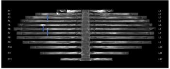
“The significant difference in the scores of fissure visualization observed in the overall study group was also found when analyzing the population stratified by (body mass index) BMI with a high proportion of fissures clearly identified in 79.3% of overweight patients and 70.6% of obese patients (vs. 25% and 27.8%, respectively (in the DECT group),” wrote lead study author Martine Remy-Jardin, Ph.D., head of the Department of Thoracic Imaging at the University Hospital of Lille in Lille, France, and colleagues.
The researchers also found a significantly shorter mean CT acquisition time with PCD-CT (0.93 s vs. 3.98 s) and a 52 percent reduction in the mean volume CT dose index (4.74 mGy vs. 9.07 mGy) in comparison to DECT. The study authors emphasized that key innovations and dose modulation capabilities enhance the viability of PCD-CT in perfusion mapping.
Three Key Takeaways
1. Superior imaging quality. Photon-counting computed tomography (PCCT) offers superior lung perfusion imaging compared to dual-energy CT (DECT), particularly with sharper delineation of fissures and fewer cardiogenic motion artifacts.
2. Reduced radiation dose. The researchers noted that PCCT provided a 52 percent lower radiation dose than DECT, which is a significant advantage in reducing patient exposure to radiation during lung perfusion imaging.
3. Efficiency and Innovation: PCCT not only shortens the mean CT acquisition time significantly (0.93 seconds vs. 3.98 seconds for DECT) but also incorporates key innovations and dose modulation capabilities that enhance the viability and quality of perfusion mapping.
“The calculation of high-quality perfusion maps on dual-source EID-CT is only possible with reduced pitch values (to reduce spiral artifacts) and reduced total collimation (to reduce the influence of cross-scattered radiation). … With PCD-CT, on the other hand, technical innovations (spectral sensitivity of the detector, better sampling in the longitudinal direction of the patient) make it possible to work with wider total collimation and a higher pitch. The absence of dose modulation with EID-CT was a theoretical disadvantage due to a constant radiation dose during acquisitions whereas dose modulation was part of PCD-CT examinations,” pointed out Remy-Jardin and colleagues.
The researchers noted the CT scans were obtained at 120 kVp with a 1.5 pitch value but acknowledged the current availability of high-pitch acquisitions and that CT scans at 140 kVp offer better spectral separation.
(Editor’s note: For related content, see “Study Says Photon-Counting CT Offers Better Lung Assessment than Conventional CT,” “Study Reveals Benefits of Photon-Counting CT for Assessing Pulmonary Embolism” and “Could Virtual Non-Contrast Images from Photon-Counting CT Reduce Radiation Dosing with CCTA?”)
In regard to study limitations, the authors conceded that utilizing similar injection protocols in both cohorts did not allow assessment of lung perfusion with a lower iodine load and pointed out there was no comparison between the groups with respect to regional lung perfusion. The researchers noted the CT scans were obtained at 120 kVp with a 1.5 pitch value but acknowledged the current availability of high-pitch acquisitions and that CT scans at 140 kVp offer better spectral separation.