|Articles|December 25, 2002
R/F and angio refine C-arms and flat detectors
Advances in C-arms appeared in the portfolios of premium performance and value-oriented vendors alike. Some included the next step up from small-size flat panels optimized for cardiac imaging to those large enough for peripheral vascular imaging. Others
Advertisement
Advances in C-arms appeared in the portfolios of premium performance and value-oriented vendors alike. Some included the next step up from small-size flat panels optimized for cardiac imaging to those large enough for peripheral vascular imaging. Others provided iterative improvements in technology more commonly tied to everyday workflow.
Eastman Kodak
The Image Sensor Solutions Division of Eastman Kodak concentrates on developing and manufacturing solid-state charge-coupled device (CCD) and complementary metal oxide semiconductor (CMOS) image sensors for medical imaging. Many of these are supplied to OEM partners for integration into private-label devices.
- The KAI-4010M Interline Transfer Progressive Scan CCD for fluoroscopy is the latest entry in the company's line of real-time digital imaging sensors. The KAI-4010M joins the KAI-2000, KAI-2093, and KAI-11000 family of sensors. It is available in sample quantities in monochrome and color versions.
- The 4-million pixel KAI-4010M features a progressive scan readout, achieving high-resolution images of quickly moving objects without distortion. The CCD employs microlens technology, which increases the ability of the sensor to transform light into electrical energy.
- At a rate of 16.5 full frames per second and 30 full frames per second at 1 K x 1 K resolution, the KAI-4010M can acquire data at high speeds for faster throughput.
GE Medical Systems
Melding in-house R&D with that of recently acquired OEC Medical, GE Medical Systems has capitalized on advanced electromagnetic technology to produce a new fluoroscopic system for image-guided, minimally invasive surgery. The company also has further advanced its homegrown Revolution digital flat-panel detector technology into a large-format interventional radiology system.
- The proprietary FluoroCAT system combines 3D fluoroscopic imaging and electromagnetic surgical navigation. FluoroCAT reconstructs 3D images from standard 2D fluoroscopic images acquired with the GE OEC 9800 mobile digital imaging system during operative procedures. Because of electromagnetic technology, the FluoroCAT system provides an uninterrupted line of sight without requiring optical tracking.
- The flat-panel Innova 4100 promises interventional radiologists images with 10 times greater dynamic range and a 37% larger field-of-view than other currently available digital fluoroscopic imaging systems. Making use of the Revolution brand of digital flat-panel technology, Innova 4100 converts x-rays into digital images at the point of acquisition and across a range of exposures, maintains image clarity over a wide field-of-view, and eliminates the artifacts and distortions associated with conventional image-intensifier chains. The Innova 4100 system has received 510(k) market clearance from the FDA and is expected to be installed in more than 100 medical facilities by the end of 2003.
Philips Medical Systems
Seeking to advance its line of general radiography/fluoroscopy and interventional products, Philips this year introduced the MultiDiagnost Eleva, a universal, multifunctional x-ray system with features targeted at R/F, vascular, and interventional radiology. The company also showcased its next-generation flat-detector technology for vascular procedures, as well as software reconstructions of calcified plaque and presurgical planning for vertebroplasty.
- The MultiDiagnost Eleva can be customized to incorporate users' preferences for performing specific types of examinations and for reviewing patients' studies. The system also includes an anticollision system, which navigates close to the viewing area by sensing the periphery of the patient. The system features 180º isocentric C-arm rotation and is equipped with DoseWise, a dose management feature that controls the radiation dose delivered to patients, as well as Grid Controlled Fluoroscopy and Intelligent Exposure, which optimize fluoroscopic and exposure parameters. This is one of the first products to include ViewForum, postprocessing software that integrates viewing and processing and customizes reporting.
- A rectangular flat detector that increases flexibility and reduces pixel size is being developed for integration into Philips' Integris Allura. The system, designed for diagnostic and interventional angiography, was released in 2001. Also in development is software aimed at providing 3D reconstructions of calcified plaque. Algorithms will identify and measure the amount of atherosclerotic calcification lining arteries. Other work-in-progress software is being written to depict 3D bony structures and implants as a prelude to vertebroplasty.
Shimadzu Medical Systems
As part of its goal of using advanced technology to simplify operations and maximize the use of space, Shimadzu has introduced C-arm innovations, a vascular digital subtraction system, a high-end remote R/F system, and fully integrated digital R/F.
- The MH-200S ceiling-mounted C-arm offers 108 programmable positions for quicker positioning, higher throughput, and improved electronic images. The multipurpose floor-mounted MH-300 vascular/cardiac C-arm features transverse travel, which allows radiologists to image the upper extremities without moving the patient or pivoting the table. Both C-arms have rotational digital subtraction angiography that can be acquired at sweep speeds up to 60º per second.
- The Digitex Premiere vascular digital subtraction system can acquire and display in a 1024 x 1024-pixel matrix as many as 30 frames per second in single-plane mode. It also has a high-speed disk array that stores up to 40,000 images in 1024 x 1024 and 120,000 images in a 512 x 512-pixel matrix.
- The all-digital, spot-filmless remote Sonialvision R/F table system is available in the U.S. in a 90/90 configuration with a 12- or 16-inch image intensifier. The II is retractable so the table can be easily lowered for loading patients.
- The Fluoromax 300 integrated digital R/F system has a large field-of-view II and can be equipped to do peripheral runoffs, thereby establishing the system as a dual-purpose R/F and angiography room. It is available with the Digitex Pro R/F and Digitex Pro Multi systems.
Siemens Medical Systems
In developing the Axiom Artis TA for angiography and TC for cardiology, Siemens Medical Systems examined the processes of interventional procedures and designed an interventional system from the ground up. The Axiom Artis series provides CT, MR, and ultrasound images at tableside, features a new high-speed gantry, and includes a flat-panel monitor and tube attached to a ceiling-mounted arm to maximize flexibility.
- Axiom Artis can retrieve any DICOM reference image, integrate color Doppler hemodynamic assessments, present data in 3D, and postprocess MR, CT, and nuclear images on a multimodality-display workstation in the interventional room.
- The interventional system also has a gantry that can move 60º per second in any direction.
Toshiba America Medical Systems
The company that pioneered the radiological application of CCD technology is taking the next step with a work-in-progress dynamic digital flat panel.
- The amorphous selenium DynaDirect flat-panel detector, which is part of Toshiba's Ultimax multipurpose x-ray system, has a pixel size of 3.3 lp/mm. The detector allows spatial resolution of 150 microns and acquires data over a surface area of about 14 inches square. The detector, which has not yet been submitted for review to the FDA, is at least nine months away from entering the U.S. market.
Advertisement
Related Content
Advertisement


Advertisement
Advertisement
Trending on Diagnostic Imaging
1
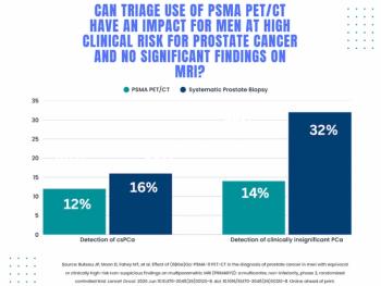
Study: PSMA PET/CT Reduces Biopsy Rate by Nearly 50 Percent for Men with Equivocal or Non-Suspicious Prostate mpMRI
2
Mammography Study: Can AI Detect Potential Breast Cancer Up to a Decade Prior to Diagnosis?
3
The Hidden Social Price of Remote Work in Radiology
4
Addressing Challenges in Radiology Reporting
5