Digital radiography teams with CR in ortho
The inherent advantages of digital radiography make it a growing favorite among radiology and other departments that use x-rays for diagnostic purposes. Speed, convenience, and quick review of images give DR an edge over rival computed radiography. Yet CR can perform some exams that are still beyond the scope of DR. This give and take is apparent in the orthopedics outpatient clinic at Strong Memorial Hospital in Rochester, NY, which manages between 250 and 300 patients per day using three DR and five CR systems.
The inherent advantages of digital radiography make it a growing favorite among radiology and other departments that use x-rays for diagnostic purposes. Speed, convenience, and quick review of images give DR an edge over rival computed radiography. Yet CR can perform some exams that are still beyond the scope of DR. This give and take is apparent in the orthopedics outpatient clinic at Strong Memorial Hospital in Rochester, NY, which manages between 250 and 300 patients per day using three DR and five CR systems.
"Technologists will try to wait for the DR room, because its throughput and efficiency help us maintain our busy work schedules," said Cindy Redmond, supervisor of imaging sciences at Strong's Clinton Crossings outpatient imaging center.
But not every patient is a DR candidate. Foot and ankle studies, which are best performed in a weight-bearing position, are typically managed with CR. For these patients, the flexibility of a cassette that patients can stand on takes precedence. But that's not to say that DR will be excluded forever.
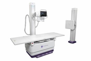
In October 2005, Strong installed a Kodak DR 7500, equipped with separate wall stand and table-based detectors that can handle a broad array of cases. Company engineers are getting feedback from the orthopedic staff on ways to improve the design. One area of interest is "standing feet" studies. As a beta site for the DR 7500, the hospital is a valued source for such information.
Strong's orthopedics department uses the DR 7500 for neonatal to geriatric patients, Redmond said. The wall stand detector accommodates three-axis movement that rotates from portrait to landscape, tilting horizontally to vertically and swinging laterally 45 degrees in either direction. A retractable bucky extends for wheelchair and table projections. Most table exposures are accomplished with a table detector that stays aligned with the overhead tube along the length of the table and can be pulled out to accommodate extremity exams. The DR 7500 is a 400-speed system, which means less radiation for patients.
"We dropped the dose by almost half compared to CR," she said.
DR is a natural for most studies, particularly those that require multiple exposures, she said. Built-in efficiencies include image display in seven to 10 seconds, immediate review and transmission to PACS, and the inherent advantage of not having to carry CR cassettes to a central reader.
"You have to take into account the time needed for a skeletal survey involving 16 or more images," Redmond said. "Add up the time involved in seven to 10 seconds per exposure to a minute per cassette, and you can see how much time we save."
Yet the staff at Strong wouldn't choose to be without CR. The flexibility of tucking a cassette under a badly injured body part makes up for delays and inconvenience. For now, side-by-side CR and DR allows ortho to handle just about any radiographic challenge.
Newsletter
Stay at the forefront of radiology with the Diagnostic Imaging newsletter, delivering the latest news, clinical insights, and imaging advancements for today’s radiologists.


