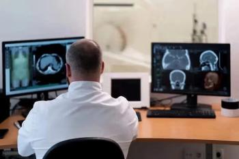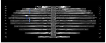
The use of adjunctive CT bone reading software in women with breast cancer detected between 8 to 22 percent more metastatic lesions for reviewing radiologists and reduced CT interpretation time by 50 percent or more, according to a new study.
Case History: 15-month-old with focal seizure, developmental delay, disproportionate increase in size of head.
Case History: A 15- month-old male patient brought by his parents with complains of one episode of focal seizure involving right side of body, developmental delay and disproportional increase in size of head (left>right). Birth history was normal: full term, vaginal delivery and unremarkable. His laboratory investigations were within normal limits.
[[{"type":"media","view_mode":"media_crop","fid":"24375","attributes":{"alt":"","class":"media-image","id":"media_crop_1150791928917","media_crop_h":"0","media_crop_image_style":"-1","media_crop_instance":"2093","media_crop_rotate":"0","media_crop_scale_h":"0","media_crop_scale_w":"0","media_crop_w":"0","media_crop_x":"0","media_crop_y":"0","title":" ","typeof":"foaf:Image"}}]]
Figure 1. T2W axial images showed disproportionate enlargement of left cerebral hemisphere with shallow sulci and enlarged gyri as compared to right side. There was enlarged lateral ventricle with colpocephaly with contralateral displacement of posterior falx and occipital lobe (arrow).
[[{"type":"media","view_mode":"media_crop","fid":"24376","attributes":{"alt":"","class":"media-image","id":"media_crop_5736737515692","media_crop_h":"0","media_crop_image_style":"-1","media_crop_instance":"2094","media_crop_rotate":"0","media_crop_scale_h":"0","media_crop_scale_w":"0","media_crop_w":"0","media_crop_x":"0","media_crop_y":"0","title":" ","typeof":"foaf:Image"}}]]
Figure 2. T1W axial images showed disproportionate enlargement of left cerebral hemisphere with shallow sulci and enlarged gyri as compared to right side. There was enlarged lateral ventricle with colpocephaly with contralateral displacement of posterior falx and occipital lobe (arrow).
[[{"type":"media","view_mode":"media_crop","fid":"24378","attributes":{"alt":"","class":"media-image","id":"media_crop_6923260001300","media_crop_h":"0","media_crop_image_style":"-1","media_crop_instance":"2096","media_crop_rotate":"0","media_crop_scale_h":"0","media_crop_scale_w":"0","media_crop_w":"0","media_crop_x":"0","media_crop_y":"0","title":" ","typeof":"foaf:Image"}}]]
Figure 3. FLAIR axial images showed disproportionate enlargement of left cerebral hemisphere with shallow sulci and enlarged gyri as compared to right side. There was enlarged lateral ventricle with colpocephaly with contralateral displacement of posterior falx and occipital lobe.
[[{"type":"media","view_mode":"media_crop","fid":"24379","attributes":{"alt":"","class":"media-image","id":"media_crop_4310582782898","media_crop_h":"0","media_crop_image_style":"-1","media_crop_instance":"2097","media_crop_rotate":"0","media_crop_scale_h":"0","media_crop_scale_w":"0","media_crop_w":"0","media_crop_x":"0","media_crop_y":"0","title":" ","typeof":"foaf:Image"}}]]
Figure 4. Gradient echo (GRE) and diffusion weighted (DWI) axial images showed similar findings as mentioned previously on T2W, T1W and FLAIR images, and no evidence of abnormal signal intensity of calcification or hemorrhage in GRE and abnormal restricted diffusion in DWI.
[[{"type":"media","view_mode":"media_crop","fid":"24380","attributes":{"alt":"","class":"media-image","id":"media_crop_3582232027744","media_crop_h":"0","media_crop_image_style":"-1","media_crop_instance":"2098","media_crop_rotate":"0","media_crop_scale_h":"0","media_crop_scale_w":"0","media_crop_w":"0","media_crop_x":"0","media_crop_y":"0","title":" ","typeof":"foaf:Image"}}]]
Figure 5. T2W sagittal images showed normally developed corpus callosum and brain stem.
MRI of the brain was performed using T2W, T1W, FLAIR sequences in 1.5 Tesla Siemens Magnetom Essenza.
On T2W, T1W and FLAIR weighted images: Disproportionate enlargement of left cerebral hemisphere with shallow sulci and enlarged gyri as compared to right side. There was enlarged lateral ventricle with colpocephaly with contralateral displacement of posterior falx and occipital lobe. However, there was no evidence of abnormal calcification or associated developmental venous anomalies.
Diagnosis: Left sided hemimegelencephaly
Discussion: Hemimegalencephaly is a rare congenital disorder of cortical malformation with hamartomatous overgrowth on all or a part of a cerebral hemisphere. This results from either increased proliferation or decreased apoptosis (or both) of developing neurons. The majority (90 percent) of patients is presented with focal and generalized infantile spasms and developmental delay, hemiparesis and hemianopia are usually also evident.
Hemimegalencephaly is divided into three forms:
1. Isolated form
2. Syndromic form: associated with a variety of syndromes typically including hemicorporal hypertrophy of the ipsilateral part of the body. Such associated syndromes include:
• Epidermal nevus syndrome
• Klippel-Trenaunay syndrome
• McCune-Albright syndrome
• Proteus syndrome
• Unilateral hypomelanosis of Ito
• Neurofibromatosis type 1 (NF1): rare
• Tuberous sclerosis: rare
3. Total hemimegalencephaly: hemihypertrophy also involves the brainstem and cerebellum
All modalities able to image the brain (ultrasound, CT and MRI) will be able to more or less identify the key features, although MRI will give the best imaging. Typical MRI features of the affected hemisphere include:
1. Increased lateral ventricle size, sometimes may be small
2. Shallow sulci enlarged gyri
3. Enlarged/thickened calvaria 4. Contralateral displacement of the posterior falx
5. White matter calcification
6. Associated with developmental venous anomalies (DVAs)
7. The cortex of the involved hemisphere may be normal, but often demonstrates polymicrogyria/ lissencephaly/agyria /pachygyria and grey matter heterotopia
Differential diagnoses include:
• Enlarged hemisphere
• Gliomatosis cerebri
• Small hemisphere, making the normal hemisphere appear large
• Rasmussen encephalitis
• Dyke-Davidoff-Masson syndrome
• Sturge-Weber syndrome
• Other neuronal migration anomalies without overgrowth
• Polymicrogyria/ lissencephaly/agyria/pachygyria
References
1. Broumandi DD, Hayward UM, Benzian JM, et al. Best cases from the AFIP. RadioGraphics 2004;24:843–848.
2. Abdel Razek AA, Kandell AY, Elsorogy LG. Disorders of cortical formation: MR imaging features. AJNR Am J Neuroradiol 2009;30:4–11.
3. Shrovan SD, Andermann F, Guerrini R, eds. The Causes of Epilepsy, Common and Uncommon Causes in Adults and Children. Cambridge: Cambridge University Press. 2011.