Advertisement
Advertisement
Trending on Diagnostic Imaging
1
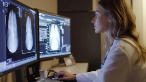
FDA Clears AI-Powered Triage Platform for Digital Breast Tomosynthesis
2
Leading Breast Radiologists Discuss the Recent Lancet Study on AI and Interval Breast Cancer
3
Is AI Better Than Neuroradiologists at Evaluating Aneurysm Growth on CTA and MRA Scans?
4
Radiology Roundup of New FDA Clearances — February 1 — February 7
5