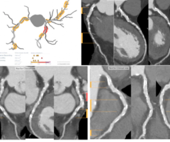
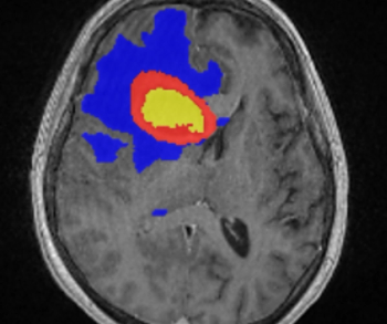
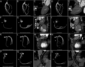
Catch up on the top radiology content of the past week.

For patients undergoing stereotactic body radiotherapy (SBRT) for prostate cancer, the acute genitourinary (GU) toxicity rate associated with the procedure was 19 percent lower with magnetic resonance imaging (MRI) guidance in comparison to computed tomography (CT) guidance, according to new research presented recently at the American Society for Radiation Oncology (ASTRO) Annual Meeting.

The automated head-to-toe shielding device, which reportedly blocks over 90 percent of radiation scatter to interventional radiologists and other clinicians, is now compatible with Siemens Artis fluoroscopy C-arm systems.

In separate test sets of Israeli women and United States women who had either ductal carcinoma in situ or invasive breast cancer, emerging artificial intelligence (AI) algorithms achieved an area under the curve (AOC) of 88 percent and 80 percent, respectively, for malignancy detection.

Catch up on the top AI-related radiology content of the past month.

The AccE Glass-Free Detector reportedly combines high-resolution capability with a variety of benefits including improved functionality, enhanced visibility, and a lightweight design.

In a new study looking at pre-operative assessment of low-grade endometrial cancer, researchers found that while magnetic resonance imaging (MRI) had 20 percent higher specificity than transvaginal ultrasound for deep myometrial invasion, there was no difference in sensitivity.

In a recent video interview from the American Society for Radiation Oncology (ASTRO) Annual Meeting, Benjamin Lowentritt, MD discussed the challenges of conventional imaging in diagnosing prostate cancer recurrence and the potential of an emerging high affinity, radiohybrid prostate-specific membrane antigen/positron emission tomography (PSMA/PET) imaging agent.

Five weeks of radiation therapy is just as effective as eight weeks of radiation treatment for men with high-risk prostate cancer, according to new research presented at the American Society for Radiation Oncology (ASTRO) Annual Meeting.

Rads will always have to deal with forces outside of their control, but it’s how we address those forces that makes us the type of radiologist we can be proud of.

The retrospective study of patients 21 years of age or younger found that a deep learning algorithm and use of the American College of Radiology’s Thyroid Imaging Reporting and Data System (TI-RADS) both had more than a 26 percent greater sensitivity for differentiating thyroid nodules on ultrasound in comparison to radiologist assessment.

For National Mammography Day, Diagnostic Imaging offers a closer look at emerging news, current insights and recent research on mammography.

For physicians performing radiotherapy treatment of soft tissue tumors in the head and neck, the MRCAT Head and Neck offers an artificial intelligence (AI) application that allows the use of magnetic resonance imaging (MRI) as the primary or sole imaging for procedure planning.

Plaque Analysis and RoadMap Analysis, two artificial intelligence (AI)-enabled assessment products, may enhance clinical evaluation of coronary artery disease (CAD) on cardiac computed tomography angiography (CCTA).

While facilitating interdisciplinary colloaboration is a central objective with enterprise imaging platforms, this author says implementation of these platforms must ensure optimal patient privacy in sensitive cases involving abuse, assault and domestic violence.

In a recent letter to U.S. Rep. Rosa DeLauro (D-CT), the Food and Drug Administration (FDA) said a final rule on amendments to the Mammography Quality Standards Act (MQSA), including an oft-delayed national standard for breast density notification in mammography reporting, may be published in the next couple of months.

The inclusion of digital bismuth germanate (BGO) detector material with the Omni Legend system reportedly more than doubles the sensitivity of older PET/CT devices, improves scan times, and enhances the detection of small lesions.

Out of 1,600 patients who had surgery for colorectal cancer, nearly 15 percent had pulmonary metastases within 15.4 months and higher-risk patients experienced lung metastases within three months, according to new research presented at the Scientific Forum of the American College of Surgeons Clinical Congress.

In a world where “gaming supplements” are available for those obsessed with video games, could radiologists be the next target for snake oil “energy” concoctions of sugar and caffeine?

Catch up on the top radiology content of the past week.

In a recent video interview, Andrew Trout, MD discussed key attributes and efficiencies in utilizing the Discovery MI Gen 2 digital positron emission tomography/computed tomography (PET/CT) system at the Cincinnati Children’s Hospital Medical Center.

A multi-year agreement with a large Chile-based supplier of raw iodine is part of GE Healthcare’s commitment to increase the production of iodinated contrast media, commonly used in computed tomography imaging, by 30 million annually in 2025.

An emerging radiomics model, derived from 68Ga-PSMA-11 PET, reportedly offers a superior area under the curve (AUC) and higher sensitivity than radiologist assessment in detecting intraprostatic lesions via positron emission tomography/computed tomography (PET/CT) in patients with prostate cancer.

As literature continues to emerge about the potential of contrast-enhanced digital mammography (CEDM), this author reviews clinical and practical benefits of the modality and pertinent steps for adding it to your diagnostic armamentarium.

In a large retrospective review of over four million Medicare claims, researchers found that Black women were 16 percent less likely to have access to digital breast tomosynthesis than White women.

While current consensus guidelines do not recommend the use of brain MRI screening in patients with breast cancer, a new study shows significantly elevated risks for the development of central nervous system metastasis in patients with inflammatory breast cancer.

While artificial intelligence (AI) models have been acknowledged for aiding imaging analysis or facilitating workflow enhancements, this author envisions AI as a potential workstation conceierge capable of turning common venting and gripes into actionable items for significant improvements.

Incorporating artificial intelligence (AI)-based technology, Neosoma HGG reportedly demonstrated a 95.5 percent accuracy rate in measuring brain tumor volume on brain magnetic resonance imaging (MRI) scans at various points during the treatment of patients with high-grade gliomas.

Catch up on the top radiology content of the past week.