


Technology
Latest News
Advertisement
Latest Videos


Advertisement
More News
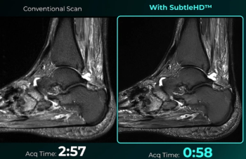
Enabling up to 80 percent faster MRI scan times, the SubtleHD software is included in the newly launched AI software suite Subtle-Elite.

Catch up on the top radiology content of the past week.

In a video interview from the International Stroke Conference (ISC), Jeremy Heit, M.D., Ph.D., discussed new research revealing over 90 percent sensitivity and specificity rates for AI detection of subdural hematomas on non-contrast-enhanced head CTs.
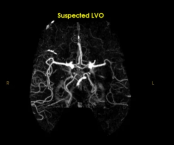
The Rapid LVO AI software detected 33 percent more cases of large vessel occlusion (LVO) on computed tomography angiography (CTA) than Viz LVO AI software, according to a new comparative study presented at the International Stroke Conference (ISC).
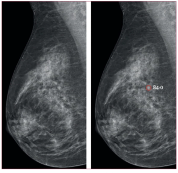
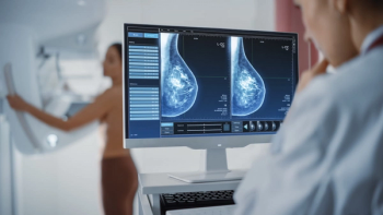
Use of the mammography AI software had a nearly equivalent false positive rate as unassisted radiologist interpretation and resulted in a 44 percent reduction in screen reading workload, according to findings from a randomized controlled trial involving over 105,000 women.
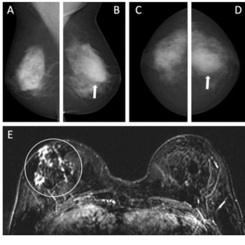
For women with intermediate risk and a personal history of breast cancer, an emerging AI system offered an 81 percent AUC for breast cancer detection, according to new research.
The artificial intelligence (AI)-enabled software Lumina 3D reportedly provides reconstructions of computed tomography angiography (CTA) images of the head and neck in minutes.
Catch up on the top radiology content of the past week.

Catch up on the most well-viewed video interviews from Diagnostic Imaging in January 2025.
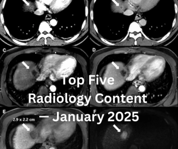
Catch up on the most-well viewed radiology content in January 2025.
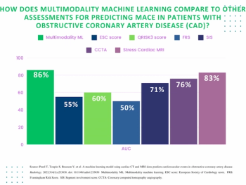
For patients with newly diagnosed obstructive coronary artery disease (CAD), a multimodal machine learning model offered an 86 percent AUC for predicting MACE, which was 10 percent higher than CCTA alone and over 35 percent higher than the Framingham Risk Score.
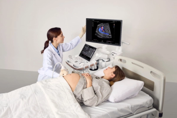
Designed for advanced OB-GYN applications, the Samsung Z20 ultrasound platform reportedly provides a combination of artificial intelligence (AI) tools and enhanced ergonomics.
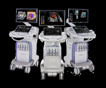
An array of AI-powered features and automated tools may lead to improved workflow and image quality with the Voluson Expert 22, 20 and 18 ultrasound systems.

Catch up on the top AI-related news and research in radiology over the past month.
Catch up on the top radiology content of the past week.
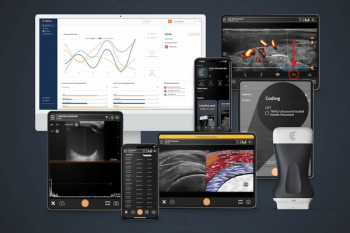
The latest ultrasound software update from Clarius includes new artificial intelligence (AI) capabilities, coding assistance and a variety of advances tailored to different fields of medicine.
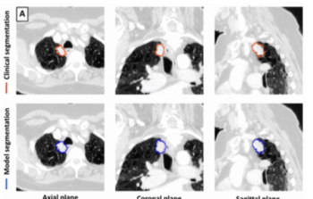
Emerging research suggests that a deep learning model may offer 92 percent sensitivity in lung tumor detection on CT scans and up to a 59 percent reduction in tumor segmentation time.
In the second part of a two-part interview, Nina Kottler, M.D., says the transparency emphasis of the recent FDA guidance on AI-enabled software is welcome but needs to go beyond additional documentation to clarify how adjunctive AI is making its decisions.
Catch up on the top radiology content of the past week.
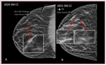
In sequential breast cancer screening with digital breast tomosynthesis (DBT), true positive examinations had more than double the AI case score of true negative examinations and the highest positive AI score changes from previous exams, according to new research.
In the first part of a two-part interview, Nina Kottler, M.D. offers insights and perspective on the recently issued guidance from the FDA on AI-enabled devices and how it may impact developers in the radiology field.
Catch up on the top radiology content of the past week.
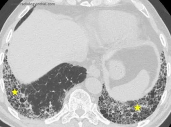
The IQ-UIP AI software may bolster computed tomography diagnosis of usual interstitial pneumonia (UIP), which is reportedly misdiagnosed in over 50 percent of cases.
In a study of over 463,000 women who had screening mammography exams, adjunctive AI led to a 17.6 percent higher detection rate for breast cancer and a three percent increase in positive predictive value for recalls.

In a recent interview, Arlene Sussman, M.D., discussed her experience in leading vRad’s teleradiology breast imaging service, how to foster personalized care in breast cancer screening, utilizing AI to help mitigate daunting worklists and improving access to subspecialty care.
Advertisement
Advertisement
Trending on Diagnostic Imaging
1
AI in Radiology: Emerging Trends, Current Obstacles and Future Directions
2
Canon Medical Unveils Advances in AI-Enabled CT Software
3
Study Says Use of 18F-flotufolastat PET/MRI May Reduce Prostate Biopsies by More Than 50 Percent
4
FDA Clears AI-Enabled Ultrasound for High-Volume Clinical Settings
5