


Opinion|Videos|September 4, 2024
Insights into the Benefit of PSMA PET Imaging Alongside Other Conventional Imaging Modalities in Prostate Cancer Care
Author(s)Jaideep S. Sohi, MD, Jeremie Calais, MD
Key opinion leaders analyze the advantages of integrating PSMA PET imaging with traditional imaging techniques to enhance prostate cancer management and treatment strategies.
Advertisement
Video content above is prompted by the following:
- From the perspective of patient care and outcomes, what potential advantages do you believe PSMA PET imaging might offer over other conventional imaging methods used in prostate cancer management?
Advertisement
Related to this article
While the allure of partnership or a hierarchical promotion system for those who paid their dues once sounded promising, the current reality amid declining reimbursements, thinning practice margins and private equity acquisitions is going after the gig with the most optimal lifestyle and paycheck.

Catch up on the top radiology content of the past week.

In the third of a three-part podcast episode, Wendie Berg, MD, PhD, FACR, FSBI, Manisha Bahl, MD, and Amy Patel, MD discuss recent developments with autonomous AI in breast imaging and share their perspective on the overall potential of AI to help mitigate the radiologist shortage.

In a recent interview, Amir Iravani, MD, FRACP, discussed the radiotherapeutic agent 177Lu-edotreotide and new dosimetry data from the COMPETE trial that was recently presented at the ASTRO conference.

While AI offers the lure of efficiency and cleaner writing in scientific research, maintaining the principles of authorship and intellectual engagement is perhaps more important than ever before.
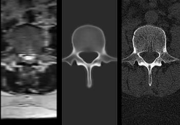
The cloud-based NextMR platform reportedly provides automated 3D bone images of conventional spine MRI images in less than 60 seconds.
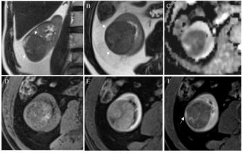
Incorporation of pseudocapsule presence into a modified Clear Cell Likelihood Score (m-ccLS) was associated with a greater than 11 percent higher AUC in differentiating benign and malignant renal masses on mpMRI, according to a new study.
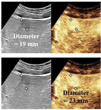
For patients with hepatocellular carcinoma (HCC), perfusion beyond tumor margin on contrast-enhanced ultrasound that exceeds gray-scale ultrasound tumor measurement by 5 mm is associated with a 76 percent higher risk of worse recurrence-free survival, according to newly published research.
Advertisement
Advertisement
Trending on Diagnostic Imaging
1
FDA Clears AI-Powered Platform for Converting Spine MRIs to 3D Visualizations
2
Diagnostic Imaging’s Weekly Scan: October 4 — October 10
3
The Reading Room Podcast: Current Perspectives, Challenges and Possibilities with AI in Breast Imaging, Part 3
4
What New Dosimetry Data Reveals About 177Lu-Edotreotide for GEP-NET Treatment
5