In the latest episode of her “Breast Imaging in Focus” series, Manisha Bahl, MD, MPH, FSBI, offer pertinent takeaways for breast radiologists from recently issued guidelines for addressing proliferative breast lesions with atypia and lobular carcinoma in situ (LCIS).
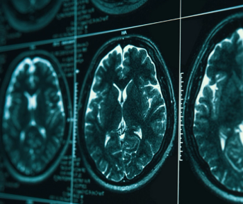
In a study involving over 2,800 people who had brain MRI scans, researchers found that people in the most disadvantaged neighborhoods had 37 percent higher white matter hyperintensity volume (WMHV) on average in comparison to people in the least disadvantaged areas.
In the second of a two-part interview, Kalpana Kanal, PhD, and Mahadevappa Mahesh, MS, PhD, FAPPM, FACR, discussed future directions for research on CT radiation dosing, including the potential of indication-specific diagnostic reference levels (DRLs).
Amid the explosion of imaging volume, an emerging silver lining is increasing recognition from referring clinicians of the time and value radiologists provide to facilitate patient care.
In a recent episode of our Molecular Imaging in Focus series, Thomas Hope, MD, discussed challenges with conventional imaging for gliomas and the potential of the newly FDA-approved PET agent floretyrosine F18 (18F-FET) for differentiating between glioma progression and treatment-related effects.
The presence of vascular hyperintensity on T2 FLAIR MRI was associated with nearly double the odds of cerebral edema, according to newly published research involving 855 patients with acute ischemic stroke.
In a recent interview, Stamatia Destounis, MD, FACR, shared her perspective on recent changes to the BI-RADS system, offering insights on contrast-enhanced mammography, reporting changes for breast MRI and ultrasound, and the importance of auditing for BI-RADS category 3 assessments.