
As I write this column, the Obama administration and the Congress are intensively discussing the elements of healthcare reform legislation.
Diagnostic Imaging Vol 31 No 11
With radiologists already working at record productivity levels, there is not much room left to maintain income levels by working harder.
I would like to call to order these hearings of the Senate Medicare Oversight Subcommittee's investigation into death panels.”
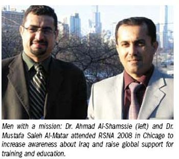
For a man whose job is to care for sick people, Dr. Ahmad Al-Shamssie's journey to work is incredibly fraught with danger. His home is about 25 km from the Baghdad Medical City complex, where he is radiology chair at the teaching hospital.
7 a.m.: IED (improvised explosive device) blocks the way to work. Many of us are late again because sections of the town are blocked. I must find an alternative route to the hospital.
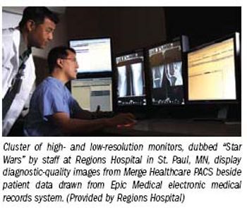
When Regions Hospital got serious about automating its patient record, administrators there decided that PACS would have to be part of it.
These are hard times for magazines, especially ones that devote themselves to medical practice.
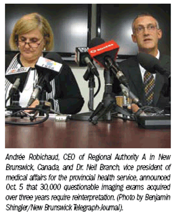
Health authorities in New Brunswick, Canada, will reexamine 30,000 imaging exams interpreted since 2007 by a radiologist based in the province’s northwestern area who is under investigation for alleged incompetence.
For some, it's sweet charity. For others, it reflects the sad state of U.S. healthcare and the general economy, but for 37 women in rural Northern California, it was an opportunity to be screened for breast cancer.
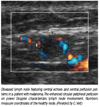
Researchers in Germany and the Netherlands have discovered ultrasound-based patterns that can accurately diagnose the presence and stage of metastases in patients with melanoma.
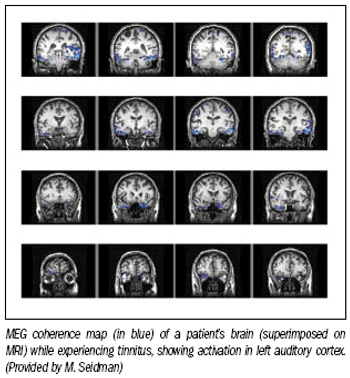
Researchers in Michigan are using magnetoencephalography of the brain to identify the source of tinnitus, a chronic ringing or buzzing in the ears or inside one's head.
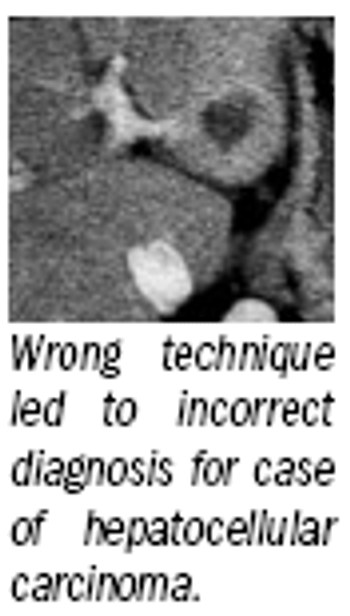
The American College of Radiology's routine announcement about a new committee for liver imaging reporting is the starting point for an ambitious plan to reinvent how radiologists diagnose and evaluate hepatocellular carcinoma.
The risks for individual imaging services are low, but radiologists should not be surprised by a visit from a representative of Medicare’s inspector general’s office to investigate the medical necessity of emergency room scans or their imaging center billing patterns.
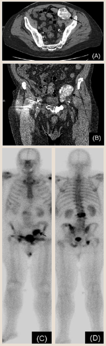
A 79-year-old man was diagnosed with prostate cancer in 1993 after a transurethral resection of the prostate (TURP). The patient had a Gleason score of 5 and was initially treated with low-dose-rate radioactive seed implants.
This month Diagnostic Imaging celebrates its 30th anniversary.
Dr. Michael E. Edwards, an interventional radiologist with Imaging Associates of North Mississippi in Corinth, and wife Sherri Edwards donated $1 million to the Society of Interventional Radiology Foundation.
Joanna Fowler, Ph.D., a major contributor to brain research and a pioneer in molecular imaging, has been awarded the National Medal of Science, the nation’s highest award for lifetime achievement in science.
Most of the general x-ray equipment currently being used in European hospitals is digital, according to a survey by InMedica, the medical branch of market research firm IMS Research.
Thanks to a near-perfect performance in a large cohort study in China, 64-slice CT angiography has shown it could replace digital subtraction angiography as the modality of choice for detecting suspected brain aneurysms at risk of spontaneous subarachnoid hemorrhage.
A large prospective trial found plaque burden and plaques involving small lumen area are as likely as classically defined vulnerable plaques to trigger a myocardial infarction.
MRI, possibly employing higher field strengths and dedicated breast coils, should be used more often for detecting ductal carcinoma in situ, according to a panel convened by the National Institutes of Health.
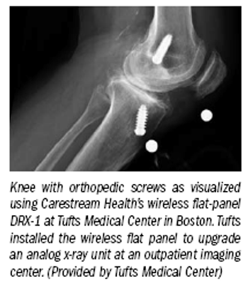
New twists on digital detectors spark vendors' hope for long-awaited commercial success.
Impact on patient safety, hospital stay lead list of vendor priorities for scanner enhancements.
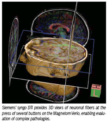
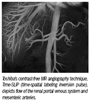
Diverse capabilities in MR reflect the corporate philosophies of individual vendors, as well as how they see opportunities in the marketplace and seek to capitalize on them
Radiologists are working approximately five more hours every week, but they also take 12 more vacation days a year, according to a survey from the American College of Radiology.
Workflow, patient comfort, image quality rise with innovative devices and applications.
Efforts to increase comfort result in speedier exams and improved image quality.
Advertisement
Advertisement
Trending on Diagnostic Imaging
1
A Closer Look at the Evolution and Emerging Insights with Amyloid and Tau PET for Dementia and Alzheimer’s Disease
2
Study Suggests FDG-PET Better than CT for Assessing Treatment Response for Metastatic Breast Cancer
3
Research Highlights from SCCT 2026
4
New Radiology Platform Facilitates Remote Access to Imaging and Collaboration
5