

How would you feel if legislators in your state approved a law requiring a specific statement in certain types of radiology reports?
How would you feel if legislators in your state approved a law requiring a specific statement in certain types of radiology reports?
Radiologists too often are either unwilling or unable to interpret screening mammography exams, so breast surgeons should learn the specialty and fill in when needed, an international speaker told a group of breast surgeons recently.
From Mike Graham, healthcare territory manager for Anthro, a vendor of digital workstation furniture:
I was surprised and a bit dismayed to read Dr. Eric Trefelner's column titled “When techs ask for ‘time out,' the end may be near” in the April 2010 edition of Diagnostic Imaging.
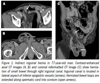
Although most hernias involving the anterior abdominal wall or groin can be diagnosed easily by inspection and palpation, imaging is the principal means of detecting internal, diaphragmatic, and other nonpalpable or unsuspected hernias.1,2
“I'm being sued for a 1 mm cancer I supposedly missed 10 years ago on a CT.”
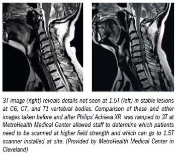
With its add-on software and higher performance coils, field-upgradeable MR scanners are the lifeblood of the imaging community. But systems with upgradeable fields are rare.
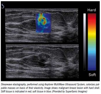
Reducing the number of breast biopsies by better classifying suspicious lesions noninvasively could improve healthcare and cut healthcare costs, laudable goals in the current era of healthcare-and economic-reform.
Breast-specific gamma imaging is highly accurate in monitoring the response of breast cancers to neoadjuvant chemotherapy, according to a study presented at the American Society of Breast Surgeons' annual meeting in Las Vegas.
A new SPECT cardiac imaging system that uses a cadmium zinc telluride-based high-speed, high-resolution camera dramatically reduces imaging time for patients while also reducing radiation exposure.
Having a pathologist onsite during ultrasound-guided thyroid biopsies can decrease the number of repeat biopsies performed due to an inadequate sample, according to a study presented at the American Roentgen Ray Society annual meeting.
PET/CT scans can confirm a suspected colorectal cancer recurrence at an early stage, helping significantly in treatment, according to a study presented at the ARRS annual meeting.
PET/CT imaging with FDG can identify when a tear in the wall of the aorta causes blood to flow between the wall's layers and forces the layers apart, according to research.
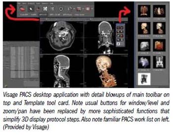
For the past 15 years, most PACS have performed the basic tasks of taking in images, archiving them, sending them to workstations for display, and hopefully not losing them.
On Jan. 24, 2009, newly sworn in President Obama uttered a bold promise: “To lower healthcare costs, cut medical errors, and improve care, we'll computerize the nation's health records in five years, saving billions of dollars in healthcare costs and countless lives.
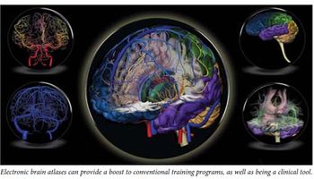
The topic of electronic brain atlases was first discussed in Diagnostic Imaging Asia Pacific almost a decade ago (see “Electronic brain atlases show value in brain studies,” June 2001, page 35). The article featured four atlases and addressed the potential of this innovation.
The adoption of digital technologies and IT in healthcare, and the consequent growth in the volume of patient data, presents hospitals with significant challenges in terms of data storage and obsolescence management.
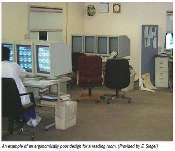
A recent study from Massachusetts General Hospital finding that radiologists suffer from poor ergonomics in the reading room put a spotlight on a relatively common problem in radiology.

The laws of physics cannot be broken-but they can be bent.