

Do you embrace challenges and change or settle and wallow in your diagnostic comfort zone?
Do you embrace challenges and change or settle and wallow in your diagnostic comfort zone?
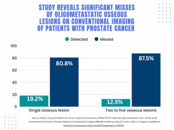
For patients with oligometastatic prostate cancer detected with PSMA PET, researchers found that nearly 78 percent of these patients had negative findings on conventional imaging, according to a study presented at the Society for Nuclear Medicine and Molecular Imaging (SNMMI) conference.
In a recent interview, Shannon Youngblood, EdD, MSRS, CNMT, RT(CT)(ARRT, shared her perspectives on advocacy for nuclear medicine technologists, scope of practice challenges, theranostics and improving access to PET imaging in rural communities.

Catch up on the top radiology content of the past week.
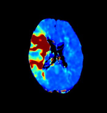
The Cone-Beam CT (CBCT) Perfusion software reportedly provides CT-quality perfusion maps during neurointerventional procedures.
Catch up on the most-well viewed radiology content in May 2026.

In a recent interview, Josh Gurewitz and Ajit Shankaranarayanan, PhD, discussed the utility of AI-powered software, including the recently FDA-cleared SubtleHD(PET), for bolstering efficiency and quality with PET imaging.
Catch up on the most well-viewed video interviews from Diagnostic Imaging in May 2026.
A randomized controlled trial found that combining Disney characters with soothing ambient elements for pediatric MRI reduced anxiety levels by 43 percent in children under 10 years of age.
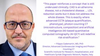
Emerging CCTA research affirms the importance of going beyond LDL reduction in the evaluation and treatment of patients with chronic coronary syndrome.
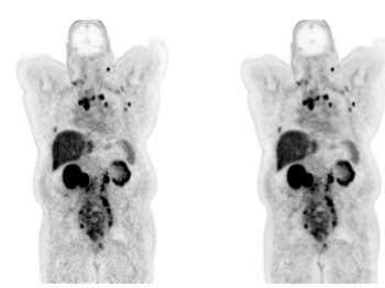
The SubtleHD(PET) software reportedly facilitates enhanced image quality, provides improved SUVmax quantitative accuracy and compatibility with all FDA-approved radiotracers.

After the U.S. Preventive Services Task Force (USPSTF) updated its biennial mammography screening recommendations to begin at 40 years of age, women in their 40s accounted for approximately 50 percent of all screening mammography exams, according to new study findings to be presented at the American Society of Clinical Oncology (ASCO) conference.

In recent interviews, Emanuel Kanal, MD, and Tobias Gilk, MRSO, MRSE, discuss new multicenter research examining pediatric MRI safety as well as key challenges and preventive measures in this patient population.
Catch up on the top AI-related news and research in radiology over the past month.
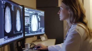
While there are pros and cons to consider, offering a direct pay service for second opinions on imaging may emerge as a viable, middleman-free alternative for radiologists.
Catch up on the top radiology content of the past week.
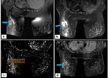
In a study of 351 women with BI-RADS 3-5 non-mass enhancement breast lesions, researchers found that multiparametric MRI offered 98.4 percent sensitivity and a 97.9 percent negative predictive value (NPV).
Catch up on a variety of new FDA clearances in radiology from the past week.

Recently published multinational research revealed that AZtrauma had a 95.6 percent AUC for detecting dislocations and a 97.5 percent AUC for detecting joint effusions.
In the latest episode of his “Interventional Radiology in Focus” series, Mina Makary, MD, discusses current and potential pre-procedure, intra-procedure and post-procedure applications of AI for interventional radiology.
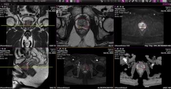
Prostate AI reportedly enhances lesion detection, reduces segmentation variability between radiologists and improves workflow efficiencies in biopsy-recommended cases.
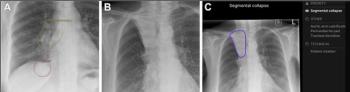
A retrospective comparison of AI platforms from seven different manufacturers for stand-alone lung cancer detection revealed sensitivity rates ranging between 20.8 percent to 77.8 percent.
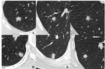
The addition of density heterogeneity on CT to a predictive model for differentiating malignant and benign pulmonary nodules led to a greater than 15 percent improvement in the model’s AUC, according to a new multicenter study.
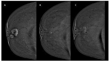
Delayed-only image acquisition for contrast-enhanced mammography after neoadjuvant chemotherapy offered a greater than 17 percent higher sensitivity rate than early-only image acquisition for detecting residual disease, according to a new study.
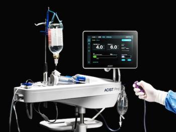
The ACIST Pro Diagnostic System, a variable-rate contrast management platform, reportedly offers real-time contrast tracking for image-guided interventional cardiology procedures and reduces set-up time by one-third in comparison to the ACIST CVI device.
A go-getter spirit, positive energy and willingness to help and learn are keys to collegial respect and propelling your career in the field.
Catch up on the top radiology content of the past week.
Researchers estimated that 17.9 million middle-aged women in the United States are overdue for breast, cervical or colorectal cancer screening, according to a new cross-sectional study.
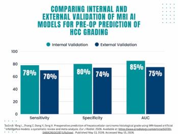
In a new meta-analysis examining the impact of MRI-based AI models for predicting high-grade hepatocellular carcinoma, researchers noted a 10 percent decline in AUC between internal validation and external validation.