

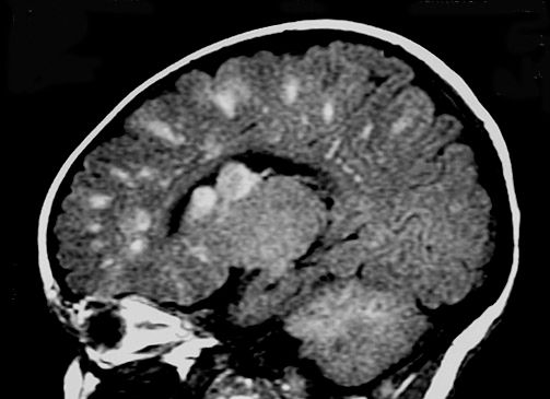


In patients with cerebral small vessel disease, brain tissue damage can occur before lesions even appear on imaging.
In patients with cerebral small vessel disease, brain tissue damage can occur before lesions even appear on imaging.

While most cases do not point to an urgent need, knowing how frequently these findings appear can help with decision-making.

Here's what to expect this week on Diagnostic Imaging.
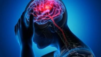
Study shows conducting MRI scans earlier in patients with mild TBI could identify which patients are likely to have the worse outcomes and when more timely intervention is necessary.
Pairing these scans with biological and clinical data can help providers with diagnosis and potential treatment.
This correlation can help providers pinpoint which patients will develop more neurological abnormalities, helping them plan interventions to improve outcomes.
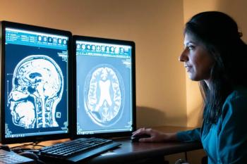
Advancements in reducing field strength are bringing MRI closer to populations unserved by the technology.
Patients receiving standard therapy and intensive therapy for high blood pressure revealed no significant biomarker differences for Alzheimer’s disease on brain images.
Here's what to expect this week on Diagnostic Imaging.
Compared with three other methods, shear wave elastography providers greater sensitivity in pinpointing remaining malignant tissue after brain tumor removal.
Images reveal ultrasound therapy can effectively open and close the blood-brain barrier, opening the door for potential new Alzheimer’s treatments.
CT Colonography & Tumor Differentiation; COVID-19 & Leukoencephalopathy; MRI, the Angiography Suite, and Acute Ischemic Stroke; Plus, DBT, African American Women, & Decreased Access
Using MRI in the angiography suite can help providers decide whether to continue with thrombectomy, to place stents, or to administer anti-thrombotic medications.
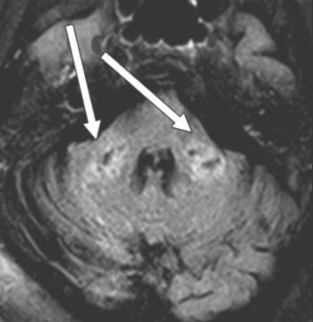
Images can show indications of rare condition that can affect white brain matter in patients who test positive for the virus.
Here's what to expect this week on Diagnostic Imaging.
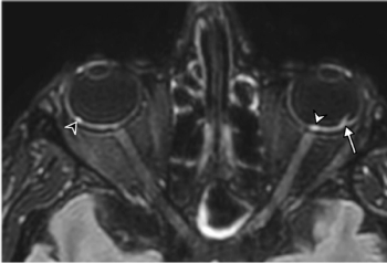
First brain MRI findings reveal dangerous COVID-19-related optical findings.
These radiopharmaceuticals are less expensive, and they offer longer half-lives.
Perivascular Spaces & Dementia; COVID-19 Loss of Smell and Taste; NAFLD & Multi-parametric CT; Plus, Point-of-Care Ultrasound in the Pandemic Era
Can you diagnose this patient with dizziness?
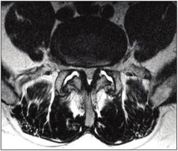
Pairing patient-reported data about pain symptoms with an MRI image can lead to better identification of pain’s origin.

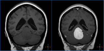
Large numbers of perivascular spaces seen in the brain are more commonly found in patients who go on to develop cognitive problems or dementia.
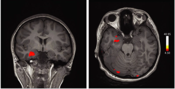
Images captured with fMRI in a case study reveal the role of the orbitofrontal cortex in patients infected with the virus who experience anosmia and ageusia.
Here's what to expect this week on Diagnostic Imaging.
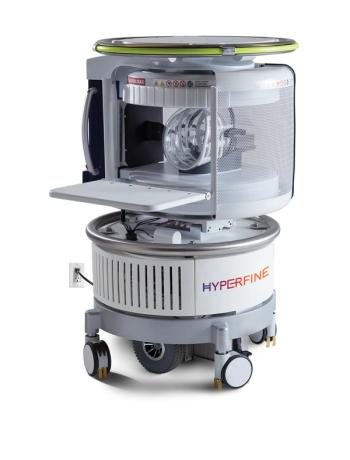
Software provides annotated and segmented brain images captured by Hyperfine’s portable MRI system.
A new magnetic resonance spectroscopy technique can accurately measure how well the mitochondria are functioning in this patient group, potentially facilitating more effective therapies.