


Magnetic resonance images showed changes in the brain, including bilateral white matter atrophy, among patients with CFS.

Magnetic resonance images showed changes in the brain, including bilateral white matter atrophy, among patients with CFS.
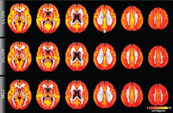
Arterial spin labeling has shown changes in the posterior cingulate cortex among patients who went on to experience cognitive decline.
Imaging can detect changes in white matter among children with reading difficulties.
Magnetic resonance imaging on patients with multiple sclerosis who use a Will balance board showed changes of the brain that affected balance and movement.
Diffusion tensor imaging detected disruptions in white matter integrity that could be linked to higher impulsivity in chronic users.

Emerging technology gives radiologists an unprecedented view of the brain.
Magnetic resonance imaging can be used to track brain growth of premature babies.
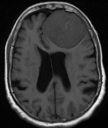
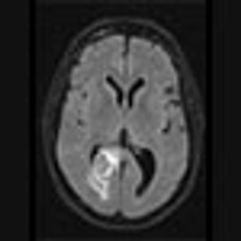
35-year-old female with prior history of breast carcinoma presented with frontal headaches.
Magnetic resonance imaging of brains of adolescents before and after the Boston marathon attack provides insight on risk factors for PTSD, study finds.
Use of MRI to visualize white matter may help clinicians predict progression of MS.
Magnetic resonance imaging may be a promising way to detect lower levels of iron in the brain, helping determine which children have ADHD.
Magnetic resonance imaging detects lower connectivity in the basal ganglia network among patients with early Parkinson’s disease.
Imaging shows signs of brain abnormalities among babies born between 32 and 37 weeks’ gestation.

Diffusion tensor imaging shows differences in the brain between males and females who sustain concussions.
Functional MRI has detected alteration in the areas of the brain that correlate with executive functioning,in boys with ADHD.
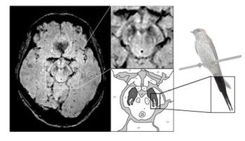
Images from 3T MRI help detect signs of Parkinson’s disease, inexpensively, and with no danger of radiation exposure.
18F-FDG PET more accurate than fMRI and may help physicians better determine the long-term recovery of patients with unresponsive wakefulness syndrome.
Diffusor tensor imaging (DTI) identifies regional white matter damage in the brains of people who sustained mild brain injuries.
Unnecessary brain scans for complaints of headache and migraine becoming more popular, but are costly for the health care system.
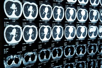
A 73-year-old male with history of a lung mass on recent chest radiograph presents with headaches.
Ultra-high-field 7-Tesla MRI of the brain detected abnormalities in the substantia nigra among subjects with Parkinson’s disease.
PET with [11C]PiB may help clinicians detect amyloid deposits in patients who have sustained traumatic brain injuries.
FDA approved GE Healthcare's radioactive diagnostic drug Vizamyl for use with PET scans in patients with Alzheimer's disease and dementia.
Insufficient evidence proving benefit of Alzheimer’s drug Amyvid behind Medicare’s ruling to limit or deny coverage.
Abnormalities in the brain among patients with back pain identified through MRI may help predict if pain will be chronic.