


Prediction occurred an average of 75.8 months before final diagnosis.
Prediction occurred an average of 75.8 months before final diagnosis.

Several lesion patterns were significantly associated with poor long-term neurocognitive and psychiatric outcomes.

Imaging the brain for iron levels may help predict disability in MS patients.
mcDESPOT magnetic resonance imaging shows changes in the myelin following mild traumatic brain injury.
Diffusion-weighted imaging after carbon monoxide poisoning may help physicians identify patients at risk for delayed neurological sequelae.
Magnetic resonance imaging shows brain pattern alterations among pre-school children with autism spectrum disorder.

DTI to monitor patients with brain metastases may help physicians determine prognosis and response to immunotherapy.
Imaging helps identify ADHD in children and may also distinguish among subtypes of the condition.
Magnetic resonance images show brain changes among adolescents who are obese.
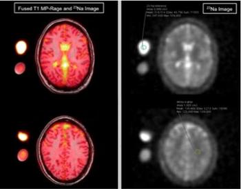
Researchers used MRI to detect higher levels of sodium in the CSF of patients who have migraines.
Imaging shows lasting brain damage among children who play high-impact sports like football.
MRIs show common structural abnormalities among patients with depression and anxiety.

MRI shows damage to brain tissue among adolescent athletes may continue after they have been cleared to return to sports.
Magnetic resonance imaging of the brain may help physicians predict patient outcomes following a cardiac arrest.
Macrocyclic GBCAs in nonenhanced T1 signal intensity pediatric brain tissue.
MRI may help physicians identify patients with Parkinson’s disease who may develop visual hallucinations.

Case History: 40-year-old female presented with history of epigastric fullness associated with epigastric and left flank pain and burning micturition.

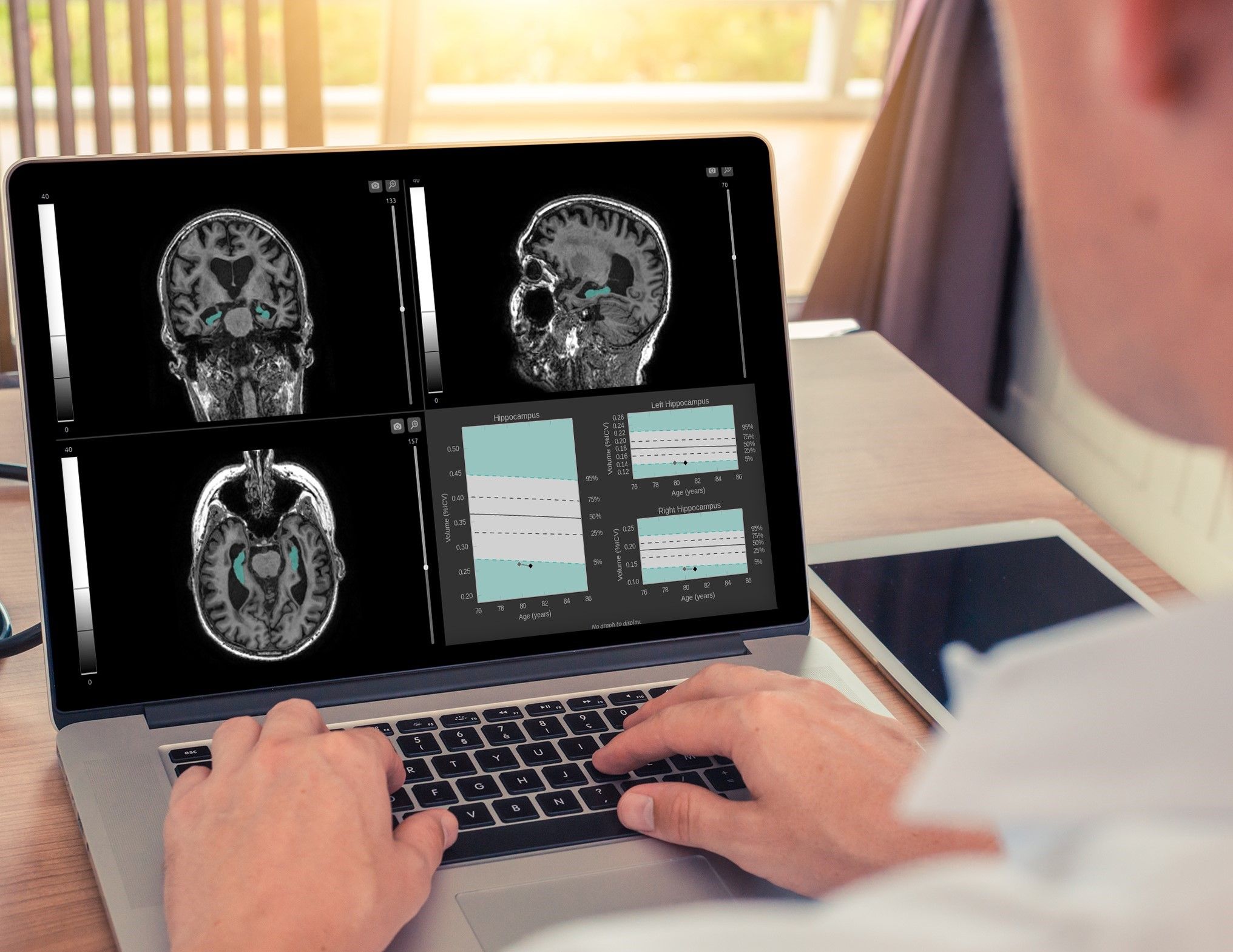
MRI detects brain serotonin levels in patients with mild cognitive decline from Alzheimer’s disease.
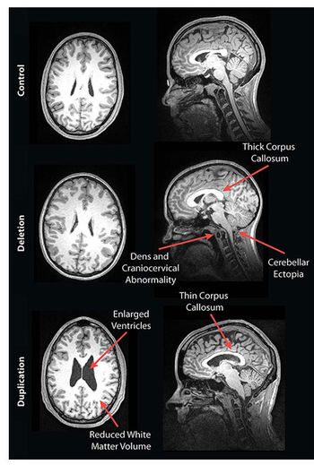
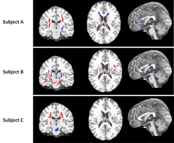
MRI has shown structural abnormalities related to cognitive function among people with genetic forms of autism.

Combined MRI can help physicians track cognitive impairment among professional fighters.
MRI may help physicians monitor Parkinson’s disease progression.
Review of MRI in the emergency department compared rates of MS as differential diagnosis and final diagnosis.

Case History: 28-year-old male presents with history of headaches.
Radiation exposure does not appear to be associated with malignant intracranial tumors among radiologic technologists.

Functional MRI may help determine which patients with depression will respond to antidepressant therapy.