Catch up on the top radiology content of the past week.
Senior Editor, Diagnostic Imaging

Catch up on the top radiology content of the past week.
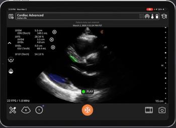
Recent research demonstrated that non-physician operators with brief training on the Accuro XV ultrasound system had a greater than 90 percent rate for obtaining diagnostically adequate images.

In a recent interview with Diagnostic Imaging, Soroush Adam Ramin, MD, discussed the enhanced 3D visualization and prognostic capability of Unfold AI, an MRI-based software platform that was recently added to Medicare Physician Fee Schedules for the West Coast and Mountain West regions.

Increased paraspinal intermuscular adipose tissue (IMAT) on whole-body MRI was associated with a 67 percent higher likelihood of hypertension, according to new research involving over 11,300 participants without preexisting conditions.

Emerging MRI findings from phase 3 research revealed that over 28 percent of patients with perianal fistulizing Crohn’s disease achieved combined fistula remission 24 weeks after initiating treatment with the IL-23 inhibitor guselkumab.

In a recent interview with Diagnostic Imaging, Wendie Berg, MD, PhD, shared her perspective on various aspects of the controversial breast cancer screening guidance issued by the American College of Physicians (ACP), including the use of supplemental imaging in women with dense breasts.

While previous research has demonstrated the capability of the PET/CT agent (89Zr)girentuximab for detecting clear cell renal cell carcinoma (ccRCC), a new analysis reveals a 98 percent positive predictive value (PPV) for detecting non-ccRCC renal cancers.

Patients with PRECISE 4 or 5 scores based on prostate MRI had over a 4.5-fold higher likelihood of biopsy progression in comparison to those with PRECISE 1-3 scores, according to a new multicenter study involving over 1,600 patients.

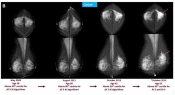
Researchers found that deep learning assessment of mammograms had an 18 percent higher accuracy than breast density in predicting future breast cancer within five years, according to a new study involving over 67,000 women.

In a recent interview with Diagnostic Imaging, Manisha Bahl, MD, MPH, SBI, offers pertinent insights on the controversial breast cancer screening guidance recently issued by the American College of Physicians (ACP).

Catch up on the top radiology content of the past week.

In a recent interview, Stamatia Destounis, MD, offered her perspective about the recent American College of Physicians (ACP) guidance on breast cancer screening and concerns over recommendations in women with dense breasts and biennial versus annual mammography screening.

Called a “star physician” by President Trump, Nicole Saphier, MD, a board-certified breast radiologist and associate professor at Weill Cornell Medical College, “is a tireless advocate for women’s health,” according to American College of Radiology (ACR) CEO Dana H. Smetherman, MD.

In an independent test set, a radiomic AI model demonstrated an 82 percent AUC for detecting pancreatic ductal adenocarcinoma on computed tomography (CT) scans at a median of nearly 16 months prior to clinical diagnosis.

In a recent interview, Matthew Hulse, a certified nuclear medicine technologist, discussed the evolution of amyloid PET imaging in managing patients with Alzheimer’s disease, the impact on PET imaging volume and the emergence of quantification platforms for monitoring treatment efficacy.

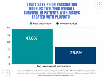
Patients who had a total tumor volume (TTV) complete response (CR) on six-week SPECT/CT imaging had a 16 percent higher survival rate at two years in comparison to those who didn’t have TTV CR, according to new prospective research.

Providing enhanced multidimensional displays of MRI and CT imaging, the Avatar Medical Vision software may facilitate improved surgical planning and intraoperative communication.

Facilitating enhanced real-time visualization for percutaneous coronary intervention (PCI) procedures, the Ultreon 3.0 optical coherence tomography platform reportedly combines high-resolution imaging with AI-powered insights on stent sizing and positioning.

In recent interviews with Diagnostic Imaging, Wendie Berg, MD, PhD, Stamatia Destounis, MD, and Manisha Bahl, MD, shared their concerns about the recent breast cancer screening guidance from the American College of Physicians.

In recent interviews, Andrew Del Gaizo, MD, Nicholas Galante, MD and David Larson, MD, MBA, shared their thoughts on the rapidly increasing imaging volume in radiology, key ramifications and possible solutions.

Catch up on the top radiology content of the past week.

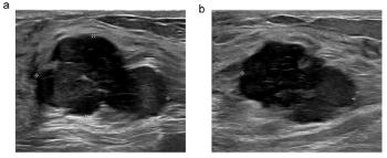
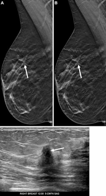
In a recent interview, Manisha Bahl, MD, discussed new breast ultrasound research findings that showed 16 percent higher specificity for breast cancer with adjunctive AI in comparison to unassisted interpretation.

AI software identified breast cancer missed by radiologists on one prior screening DBT exam in 26.8 percent of patients and on three prior screening DBT exams in 11 percent of patients, according to a study involving over 300 women that was presented at the Society of Breast Imaging conference.

In a recent interview with Diagnostic Imaging, Sujith Kalathiveetil, MD, FACC, discussed the evolution of coronary computed tomography angiography (CCTA), AI-enabled tools and the potential for future innovations with CCTA.


The Rembra CT reportedly facilitates high-throughput imaging for high-volume settings such as emergency departments and the Rembra RT and Areta RT platforms offer an extended 85 cm field of view and 4D CT to enable enhanced accuracy in tumor targeting.

PlaqueSegNet, an emerging deep learning model for coronary plaque quantification based off CCTA exams, offered a greater than 90 percent intraclass correlation coefficient agreement with intravascular ultrasound (IVUS) and expert readers across four separate datasets.

In a recent interview, Andrew Del Gaizo, MD, and Nicholas Galante, MD, shared their thoughts and perspectives on increasing imaging volume, the sunsetting of PowerScribe 360, AI hype and promise, and potential opportunities in radiology.

Facilitating real-time visualization of deep vessel structures and high-resolution surface detail within a single workflow, the hybrid intravascular ultrasound (IVUS)/optical coherence tomography (OCT) platform will be launched in the second quarter of 2026.

Catch up on the top radiology content of the past week.