


MRI plus clinical criteria helps physicians determine which stroke patients would benefit from thrombectomy.
MRI plus clinical criteria helps physicians determine which stroke patients would benefit from thrombectomy.
CHICAGO-MRI suggests that subclinical cardiac dysfunction could be indicative of early brain disease.

CHICAGO-MRI shows abnormal cerebral blood flow even if concussed athletes clinically ready to return to sport.
28-year-old male presented with sensorineural hearing loss for two months.
Using MRI to assess hippocampal grading, physicians may be able to predict Alzheimer’s disease up to seven years before dementia.

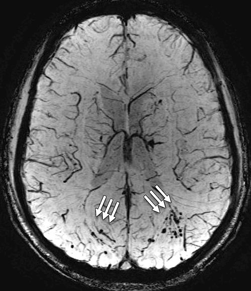
An earlier scan of a patient’s brain with traumatic brain injury is better for greater detection of microhemorrhages.

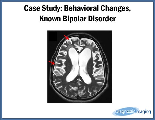
Use of MRI images may help psychiatrists determine effectiveness of antipsychotic drug treatment.

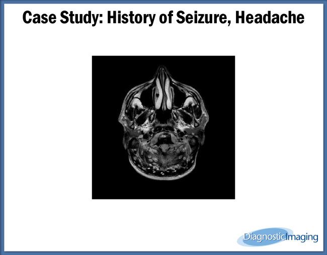
Case History: 35-year-old presents with history of seizures.
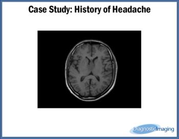
Case History: 40-year-old with history of headache.

Case History: 24-year-old presents with complaints of headache and seizures.

This Q&A series explores radiology’s role in overdiagnosis in a variety of conditions. Here, we discuss mild traumatic brain injury.

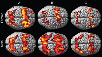
Magnetic resonance imaging shows that women have changes in the brain’s gray matter volume after stimulant addiction.
Cerebral lesions of 3 mm or smaller, detected by MRI, may increase risk of stroke and death.

Using diffusion-tensor imaging, researchers found brain changes among patients with mild traumatic brain injury who are depressed or anxious.
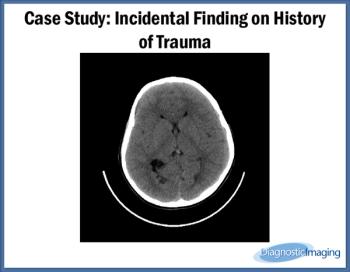
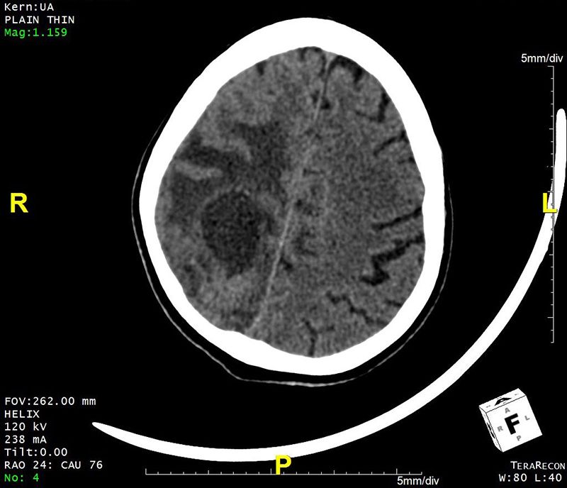
Case History: 27-year-old patient presented with history of trauma.

Neuroimaging may not be necessary for all children with sports-related concussion.

MRI may be able to detect an early marker of atypical Alzheimer’s disease.
Magnetic resonance imaging shows lower digit span scores among women with MTBI.
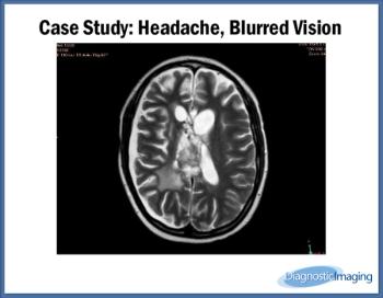
Case History: 20-year-old female with severe headache, blurred vision, vomiting.
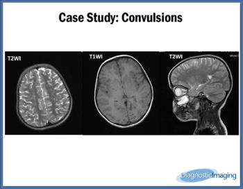
Case History: 7-year-old female with complaints of convulsions for one year.

A new, free, website provides comprehensive answers to complex questions about MRI.

Case History: 47-year-old male with headaches followed by generalized tonic-clonic convulsions.
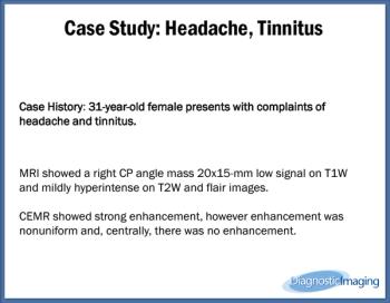
Case History: 31-year-old female presents with complaints of headache and tinnitus.

Neuroimaging with MRI shows an association between brain structure and postconcussive symptoms.

Choosing Wisely guidelines can miss important findings, neurosurgeons argue.