
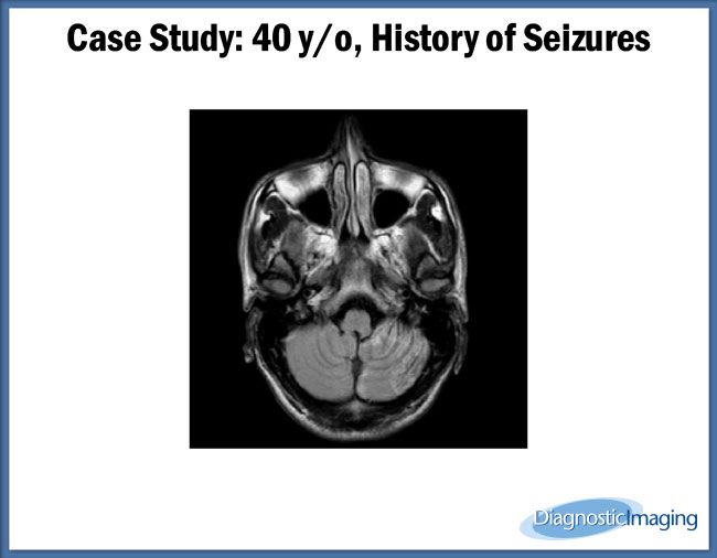


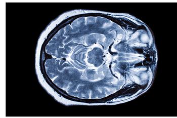
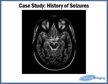
Case History: 45-year-old male with history of alcoholism with history of four seizures one day earlier.
Case History: 45-year-old male with history of alcoholism with history of four seizures one day earlier.

MRI shows exercise may help adults with mild cognitive impairment.
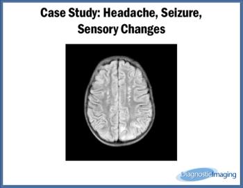
Case History: 32-year-old patient presented with complaint of headache, seizures, and sensory changes.

MRI indicates that traumatized boys and girls may have differences in insula subdivision structure.
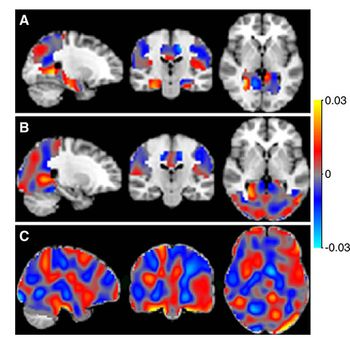
Future neuroimaging studies of psychotherapy response may focus further on individual regions in the brain.
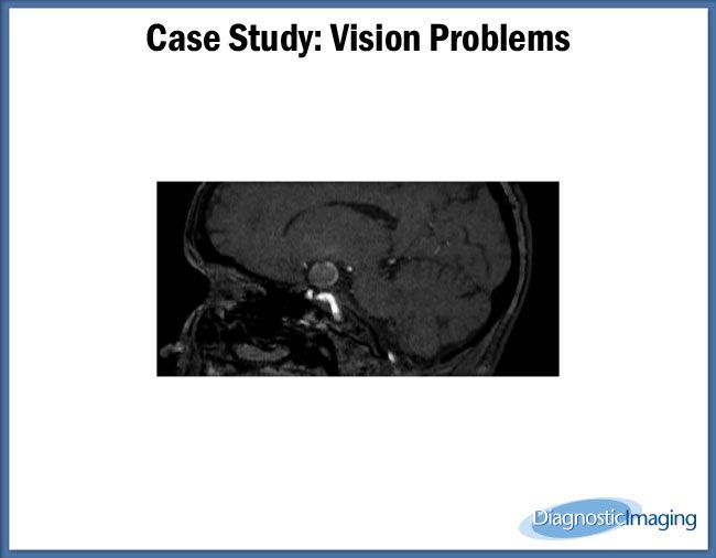
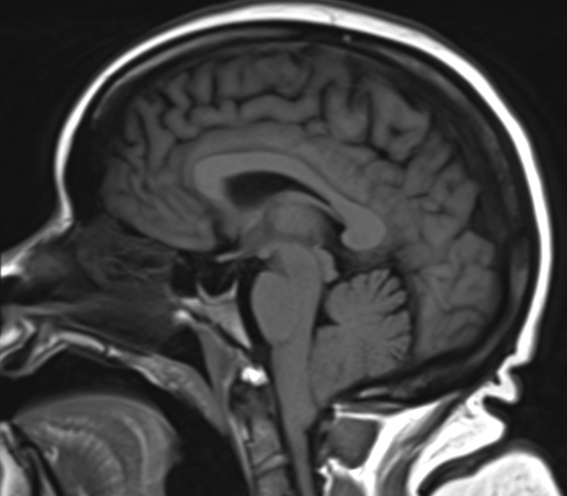
MRI images often detect incidental findings in older patients.

MRI highlights changes in brains of children with Tourette syndrome.

MRI shows brain changes in children with PTSD.

MRI shows brain changes in children who play football even if not diagnosed with concussion.
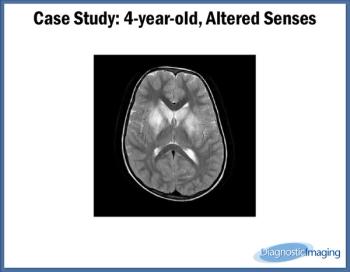
Case History: Four-year-old patient with complaints of altered sensorium.
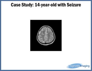
Case History: Fourteen-year-old presented with complaints of seizure.
Brain MRIs may help diagnose vascular cognitive disorder following strokes and TIAs.
Case History: 27-year-old patient presented with history of seizures.
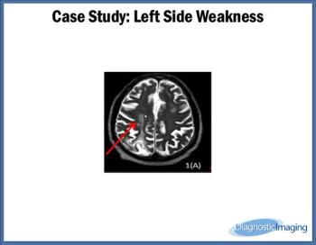
Case History: 70-year-old male with left-sided weakness and history of myocardial infarction four years prior.
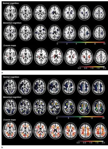
Adding arterial spin labeling to MRI may help classify and predict Alzheimer’s diagnosis and progression from subjective cognitive decline.

Functional MR imaging shows increased response in the brain after administration of low-dose methylene blue.

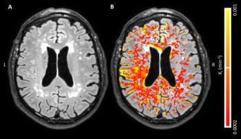
Using MRI to evaluate blood-brain barrier disruption following intracranial hemorrhage may predict severity after intervention.
Diffusion tensor imaging detected abnormalities in the brain among subjects with mild traumatic brain injury.
MRI may help detect Alzheimer’s before directly visible cerebrovascular abnormalities are present.

Use of brain imaging in the ED increased dramatically since 1994, according to a study at ACR 2016.
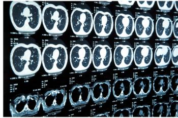
Brain CT and MRI for early stage lung cancer patients are not routinely recommended.

Radiologists detect changes in the brains of veterans who have sustained combat-related mild traumatic brain injuries.
Diffusion-tensor MRI algorithm may help physicians evaluate patients with mild traumatic brain injury and post-trauma migraines.

MRI distinguishes brain lesions, possibly supplementing existing diagnostic algorithms
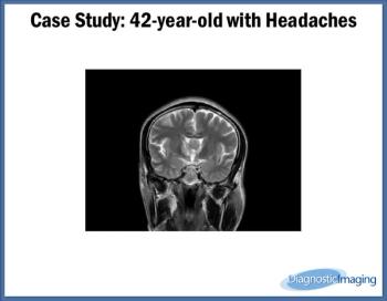
Case History: 42-year-old patient presents with complaints of headache.