|Articles|August 23, 2005
64-slice beats 16-slice CT in virtual colonoscopy
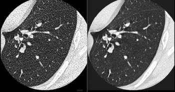
CT colonography performed on a 64-slice scanner produces superior image quality and lesion delineation compared with exams performed on a 16-slice machine. The newer technology’s faster scan time also reduces motion artifacts, according to a scientific exhibit at the European Society of Gastrointestinal and Abdominal Radiology meeting in Italy in May.
Advertisement
CT colonography performed on a 64-slice scanner produces superior image quality and lesion delineation compared with exams performed on a 16-slice machine. The newer technology's faster scan time also reduces motion artifacts, according to a scientific exhibit at the European Society of Gastrointestinal and Abdominal Radiology meeting in Italy in May.
Dr. Anno Graser and colleagues at the Ludwig-Maximilians University Munich screened 30 individuals in the prone and supine positions using a 16-slice scanner at a collimation of 0.75 mm, and 30 patients on a 64-slice system at 0.6-mm collimation.
Two independent expert readers using endoluminal views rated image quality on a five-point confidence scale. They assessed visibility, delineation of lesions, and anatomic details in four colonic segments: sigmoid, descending, transverse, and ascending.
The average exam time was cut by nearly 50% using the 64-slice protocol, from 15.5 seconds to eight seconds. The newer scanner produced no motion artifacts, while five were recorded for the 16-slice machine, a significant difference.
The mean image quality increased from 4.2 to 4.7. At p<0.05 for indication of statistical significance, the 64-slice scanner achieved a superior visualization of lesions and normal colonic mucosa, according to the study.
The Munich researchers also presented at the meeting results of a study that evaluated a prototype computer-aided detection system for CT colonography compared with two expert readers. The CAD system showed high sensitivity in detecting clinically significant polyps greater than 6 mm with acceptable false-positive rates.
Graser and colleagues scanned 105 asymptomatic patients using a 16-slice scanner at 0.75-mm collimation in the supine and prone positions without fecal tagging. Polyps less than or equal to 6 mm were classified as small, those between 7 mm and 9 mm were classified as medium, and those at 10 mm or greater were classified as large.
Readers found 98 polyps (51 small, 32 medium, and 15 large) in 42 patients. CAD detected 69 polyps (70% overall sensitivity) with an average of 3.8 false positives per data set. Average CAD running time was 4.8 minutes. Sensitivity was 51% for small, 94% for medium, and 87% for large polyps.
Investigators concluded that the short calculation time of the CAD system makes its use feasible in screening.
For more information from the Diagnostic Imaging archives:
Advertisement
Related Content


Advertisement
Advertisement
Trending on Diagnostic Imaging
1
Should There Be Greater Oversight of MRI Safety?
2
Thirteen Takeaways from New Report on AI in Health Care
3
Mammography Study: Can AI Detect Potential Breast Cancer Up to a Decade Prior to Diagnosis?
4
FDA Clears AI-Powered Software for Improving Low-Contrast CT Detection
5