"CT angiography is extremely sensitive; it is just not specific. It can show that people have coronary artery disease when blood flow to the heart is actually OK," said Dr. Henry Wagner, a professor of nuclear medicine at the Johns Hopkins School of Public Health.
In an award-winning study, Dr. Phillip Kaufmann and colleagues at University Hospital Zurich in Switzerland demonstrated that MSCT alone should not be trusted to triage low-risk cardiac patients. They prospectively studied 62 consecutive patients with suspected CAD using SPECT/CT. The 64-slice CT angiography generated only a 54% positive predictive value for 75% or greater stenosis on a per-patient basis and a 90% negative predictive value for ruling out significant disease.
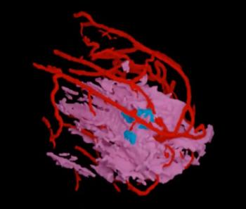
While the addition of SPECT to CTA data boosts specificity, MSCT alone is a reliable tool for screening a low- to intermediate-risk population, Kaufmann said, pointing out its high sensitivity (91%) and negative predictive value (97% for stenosis less than 50%). The accompanying image from the Kaufmann study won the Image of the Year award at the SNM meeting.
Fusion SPECT/CT may have implications for complex cardiac cases as well, according to SNM president Dr. Martin P. Sandler. Speaking at a GE-sponsored symposium, Sandler described his initial experience with the investigational GE Ventri VCT scanner. Indicative of the next generation of hybrid cameras, it features a combined 64-slice CT and SPECT camera for simultaneous attenuation correction, cardiac image acquisition, and fusion.
"It is a big problem we are about to solve here, when you get patients with complex coronary artery disease and the interventional cardiologist is limited in deciding where to do an invasive procedure or whether the patient needs to get back to surgery," Sandler said.