
|Articles|May 1, 2007
Digital mammograms shrink down to more practical size
Wavelet compression can shrink large, unwieldy digital mammography images by a factor of 100 without any loss in image quality, according to a German study presented at the European Congress of Radiology in March.
Advertisement
Wavelet compression can shrink large, unwieldy digital mammography images by a factor of 100 without any loss in image quality, according to a German study presented at the European Congress of Radiology in March. This finding has tremendous potential benefits for productivity, transmission speed for teleradiology, and PACS storage costs.
The study, which involved a standard wavelet compression algorithm, indicates there is little difference in quality between a standard size original mammogram of 33 MB and one that has been compressed to as small as 325 KB, said Dr. Reinhard Loose, head of radiology at the Klinikum Nuremberg.
Image size varies depending on modality. Large image size is particularly problematic in mammography because some health authorities, such as the FDA, currently do not allow lossy compression. Studies showing equivalent image quality could help bolster the case in favor of lossy compression, which is permitted in other modalities.
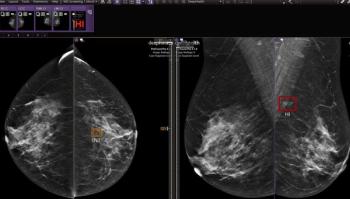
In the Nuremberg study, nine skilled radiologists with more than four years each of experience in mammography compared original and compressed digital mammography images for three patient groups: normal results, microcalcifications, and small carcinomas. The study included both computed radiography and flat-panel detector full-field digital mammography units.
The radiologists rated image quality and ranked images for both CR and FFDM. In cases that appeared to have a loss in quality, they were asked if the image was still of diagnostic quality.
The compression rate threshold was set at 100 because researchers presumed loss of quality would occur somewhere beneath that level, as is the case with other types of diagnostic imaging studies. Computed radiography of the lung, for example, shows artifacts beyond compression of 1:50.
Mammography results came as a surprise, according to Loose.
"There was no statistically significant impact on quality or visible difference in images for compression rates up to 100," he said.
Compression varies depending on the size of the matrix, and PACS should be able to automatically apply the right level according to the modality, Loose said. Brain CT images, for example, have a 512 matrix, meaning every pixel can be seen with the human eye and there is an extremely low dynamic range of 80 Hounsfield units out of 4096 stored units. Maximum compression is 1:5.
"Images can be compressed higher if they have a larger matrix size and a wide dynamic range. Hence, mammograms offer the highest compression without loss of diagnostic image quality," Loose said.
Even if radiologists played it safe by compressing mammography images by a factor of just 30, they would be able to send an image in one minute over a standard ISDN line, he said.
The researchers now plan to analyze the effect of increasing compression to the 200 level. They're also planning a randomized controlled study comparing the quality of original mammograms to ones compressed by a factor of 50.
Advertisement
Related Content
Advertisement


Advertisement
Advertisement
Trending on Diagnostic Imaging
1
What Does the Future Hold for Nuclear Medicine?
2
Top Five Radiology Content in June 2026
3
What New MRI Research Reveals About Endometrial Cancer Staging and the 2023 FIGO Staging System
4
FDA Clears New CEM Image Reconstruction and Biopsy Capabilities for Siemens Healthineers Mammography Device
5