|Articles|October 1, 2007
Glut of incidental lesions leaves little time to achieve reporting consensus
High-resolution imaging has enabled radiologists to pinpoint pathology with uncanny accuracy. The flip side of this technological boon, however, is the increasing number of incidental findings and a general lack of consensus about how to report them. Without some broad agreement, radiologists run the risk of overcalling benign lesions or undercalling malignant ones.
Advertisement
High-resolution imaging has enabled radiologists to pinpoint pathology with uncanny accuracy. The flip side of this technological boon, however, is the increasing number of incidental findings and a general lack of consensus about how to report them. Without some broad agreement, radiologists run the risk of overcalling benign lesions or undercalling malignant ones.
In 2005, experts reached consensus regarding incidental findings at ultrasound in the thyroid. But it was consensus in the broadest terms, "what we in the room could agree upon," said Dr. Franklin Tessler, chief of body imaging at the University of Alabama in Birmingham (see Overread, July, page 10). The American College of Radiology this year charged a panel with reaching consensus on unexpected findings in the kidneys, liver, adrenals, ovaries, and pancreas (see Overread, page 11).
"Although there are studies that address common benign lesions, I think the ACR panel will have difficulty finding enough peer-reviewed data for a consensus on incidental findings in the abdomen and pelvis," said Dr. Jay P. Heiken, director of abdominal imaging at Washington University in St. Louis.
A Medline search on incidental pancreatic lesions exemplifies the problems faced by expert panels. One study cautioned against conservative management (Arch Surg 2003;138:427-434) and another against relying on preoperative characteristics alone to determine the malignant potential (Am J Surg 2006 Aug;192(2):148-154). Two other studies found that pancreatic cystic lesions can be managed conservatively (Clin Gastroenterol Hepatol 2007;5:813-817 and Radiology 2006;238:912-919).
Most incidental gastrointestinal/genitourinary findings are in the liver, kidneys and adrenal glands (roughly in that order), with an increasing number in the pancreas (see "Multislice CT and 3D propel pancreatic imaging forward," page 33). The kidneys harbor the most malignant unexpected findings (see "MR imaging, CT offer answers to renal mass questions," page 45).
Dr. Alec Megibow, a luminary in GI imaging at New York University, says the single most important weapon against the overcalling of incidentalomas is comparison with any old imaging study.
"I do not find this to be universally practiced. I hope that any white paper written by the ACR committee will stress the absolute necessity of rigorously attempting to acquire prior imaging studies. I cannot tell you how many patients were rescued from biopsy or other intervention when their old studies materialized," Megibow said.
Dr. Alan Kaye, radiology chair at Bridgeport Hospital in Connecticut, sees a culture of timidity among radiologists springing from an emphasis on defensive radiology. He teaches residents that they are not merely catalogers of findings but consultants who make judgments and assessments. To bring some standardization to the reporting of incidental lung nodules, Kaye's practice adopted a series of recommendations from the Fleischner Society (Radiology 2005;237:395-400). These guidelines appear at the bottom of every report where incidental nodules are found.
Kaye and company are also modifying the ACR's RADPEER classification to better deal with incidental findings. In the RADPEER program, a radiologist can review a colleague's findings and concur to some degree (categories 1 and 2) or disagree (categories 3 and 4). Kaye's group has decided to add category 0 to ensure that colleagues assess incidental findings and don't just announce their presence.
Radiology is at a crossroads. Advanced technology has enabled many lesions to be detected and treated earlier. But the murky area between benignity and malignancy in many cases demands more research so solid consensus can be drawn. Otherwise, this will become another area in which the threat of malpractice rather than the quality of care dictates practice.
Advertisement
Related Content
Advertisement


Advertisement
Advertisement
Trending on Diagnostic Imaging
1
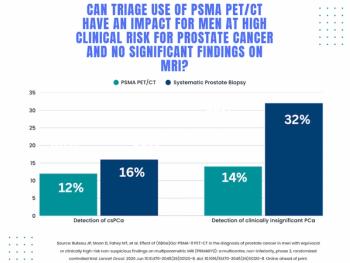
Study: PSMA PET/CT Reduces Biopsy Rate by Nearly 50 Percent for Men with Equivocal or Non-Suspicious Prostate mpMRI
2
Addressing Challenges in Radiology Reporting
3
The Hidden Social Price of Remote Work in Radiology
4
Mammography Study: Can AI Detect Potential Breast Cancer Up to a Decade Prior to Diagnosis?
5