
|Articles|October 22, 2001
PACS and ophthalmology: the eyes have it
Ophthalmology can join radiology and cardiology in the digital world, following the recent announcement of a PACS-ophthalmology solution from Agfa. The system essentially digitizes the retinal, or fundus, cameras used as diagnostic tools by
Advertisement
Ophthalmology can join radiology and cardiology in the digital world, following the recent announcement of a PACS-ophthalmology solution from Agfa.
The system essentially digitizes the retinal, or fundus, cameras used as diagnostic tools by ophthalmologists, optometrists, and other medical professionals, particularly in the detection and monitoring of diabetes.
Prior to introduction of the new modality, retinal images could only be captured on 35-mm film, a method implying darkrooms, developing chemicals, cutting and snipping, and reading cards distributed by hand or mail.
Now, the images can also be digitized, providing ophthalmologists with the same image transmission, viewing, and storage benefits previously available elsewhere only to PACS users.
"The big change in the technology for ophthalmologists is that we can supply them with backend networking, archive, and distribution of images anyplace, anytime," said John DeSantis, director of global ophthalmology at Agfa.
The system, which Agfa will show for the first time at the November American Academy of Ophthalmology meeting in New Orleans, is basically a mini-PACS, with smaller footprint and functionality features to meet a price point that fits the market.
The ophthalmology PACS system has attracted the attention of the federal government, which is interested because of the high incidence of diabetes among patients in Veterans Affairs hospitals and in the Indian Health Service.
The system is already fully implemented at the Seattle VA Medical Center and three outlying clinics networked to it, providing proof-of-concept.
A growing number of ophthalmologists have been lurking around the PACS perimeter for some time, particularly in subspecialty areas like neuro-ophthalmology.
"We use the hospital PACS whenever we order radiologic procedures on our patients," said Dr. Larry Frohman, an associate professor of ophthalmology and neuro sciences at the University of Medicine and Dentistry of New Jersey (UMDNJ), whose department of eight neuro-ophthalmologists went filmless this summer.
Ophthalmologists and radiologists at UMDNJ rotate through several hospitals, and going filmless made life simpler for the clinicians
"It would take almost an act of Congress for both of us to get in the same place at the same time to look at the films together, providing the film could even be found," Frohman said. "Now we can consult with the radiologist in near real-time."
Frohman said ophthalmologists are particularly fond of being able to view night-call images from home.
"This revolutionizes things, particularly once we all get high-speed access lines," he said. "I'm in a place that sees a tremendous amount of trauma, so it's common for residents to call in the middle of the night needing an opinion on a patient with an orbital fracture or an optic nerve contusion."
Until about a month ago, that would mean Frohman might have to get up and travel to the university just to see the x-ray. Now ophthalmologists can switch on their home computers and view the studies extemporaneously.
Advertisement
Related Content
Advertisement


Advertisement
Advertisement
Trending on Diagnostic Imaging
1
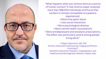
The Hidden Social Price of Remote Work in Radiology
2
Addressing Challenges in Radiology Reporting
3
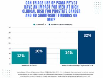
Study: PSMA PET/CT Reduces Biopsy Rate by Nearly 50 Percent for Men with Equivocal or Non-Suspicious Prostate mpMRI
4
Breast Imaging in Focus: Can GLP-1 Receptor Agonists Reduce Breast Cancer Risk? What New Research Reveals
5