
|Articles|December 1, 2006
Siemens swaps goggles for sophisticated display
Some vendors promote 3D imaging and others 3D display. Now Siemens Medical Solutions has both. The company is selling a monitor that produces volumetric images that float in space beyond its flat screen. This capability was possible in the past but only when wearing glasses with multicolored lenses or shutters that snapped open and closed 25 or more times a second.
Advertisement
Some vendors promote 3D imaging and others 3D display. Now Siemens Medical Solutions has both.
The company is selling a monitor that produces volumetric images that float in space beyond its flat screen. This capability was possible in the past but only when wearing glasses with multicolored lenses or shutters that snapped open and closed 25 or more times a second.
Siemens' 3D Stereoscopic Display System needs nothing more than a pair of interested eyes. The system was shown as a work-in-progress at the 2005 RSNA meeting and commercially released a few weeks before the 2006 meeting.
"The trick is getting different images to the different eyes," said Michael Martens, product manager for Siemens' angiography/x-ray division.
The face of the flat-screen monitor is composed of microlenses that match up with individual pixels in the 3D image. As many as eight views of the static image are projected through these microlenses. Some are perceived by one eye, some by the other eye.
When the brain puts the two images together, a stereoscopic impression is created that provides the illusion of depth. In short, Siemens' new display system mimics the way the human eye works.
Sold as an option to the Siemens' syngo Workplace, the display provides a clear view of vascular structures, such as vascular aneurysms, stenoses, and vascular malformations. It is particularly suited to data obtained using rotational angiography but is equally at home with data from any angiographic modality, including CT and MR imaging.
Interventionalists and surgeons will want to use the display to visually assess vascular structures, according to Martens, as well as the paths to reach them. The reason has to do with ease of use.
On a conventional flat-panel display, reconstructions must be rotated back and forth to get a 3D impression, he said, and to see which vessel is behind which structure.
"With the stereoscopic display, you have all that depth information right there," he said.
Outwardly, the stereoscopic monitor looks exactly like a medical-grade liquid crystal display. It costs twice as much, however. The company says that is because of the sophisticated technology that powers the system. The multiple images projected through the microlenses must be specially processed and meticulously managed to create a virtual 3D model, Martens said.
Just as the display eliminates the need for special glasses, it can be viewed by more than one observer at the same time, making the display useful in consultations or surgical planning sessions.
"As you move your head around, you see different views of the static object," Martens said.
The volumetric presentation has one drawback, however. Projecting a multifaceted image degrades that image.
"You lose about one-eighth of the resolution," he said.
This loss may be acceptable, however, considering that the operator has the option to view the data conventionally.
A single button on the stereoviewer changes the display from 2D to 3D. Press another button on the viewer, and the display returns to 2D.
"The postprocessing to create a 3D display is all done in the background," he said. "The operator only has to press the button and go for it."
Advertisement
Related Content
Advertisement


Advertisement
Advertisement
Trending on Diagnostic Imaging
1
Emerging PET Radiotracer May Enhance Detection of Small Metastases in Patients with Advanced Melanoma
2
Molecular Imaging in Focus: Key Takeaways from the 2026 SNMMI Conference with Michael Hofman, MBBS, FRACP
3
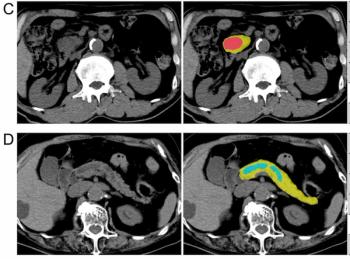
Can AI Enhance Non-Contrast and Contrast-Enhanced CT Detection of Pancreatic Cancer?
4
Five Strategies to Facilitate Technology Implementation and Alleviate Radiologist Turnover
5