Register Now: Image-Based Risk: The Next Frontier in Breast Cancer Screening
|Articles|December 1, 2003

Technique and patient prep prove key in detecting colon polyps with virtual colonoscopy

Virtual colonoscopy is gaining in clinical stature as a viable screening tool for colon cancer, with high sensitivity and specificity in detecting colonic polyps. But proper technique and good patient preparation are key to success, according to research presented Monday.


Virtual colonoscopy is gaining in clinical stature as a viable screening tool for colon cancer, with high sensitivity and specificity in detecting colonic polyps. But proper technique and good patient preparation are key to success, according to research presented Monday.

In a presentation that provided updated results from a two-year screening trial comparing virtual colonoscopy with traditional colonoscopy, interpreting radiologists relied primarily on 3D endoluminal displays, using 2D images for correlation and problem solving.

Virtual colonoscopy detected more than 90% of significant polyps in 1233 asymptomatic patients, including 92.6% of polyps 8 mm or greater. Conventional colonoscopy detected 89.5% of such polyps. The virtual technique performed best in polyps at least 6 mm in size, said Dr. J. Richard Choi, lead investigator for the study, which was conducted at several Department of Defense sites and based at Walter Reed Army Medical Center.

Technique and patient prep proved critical to the study's high success rate, Choi said. Bowel preparation included phospho-soda and dilute oral CT contrast in two divided doses. For patients in whom polyps were detected and treatment indicated, this patient prep allowed same-day or next-day performance of a colonoscopy exam.

The 3D primary reading technique also contributed to the high detection rate, Choi said.

"We found that reading in primary 3D mode was easy with the software we were using," he said. "We also found that the learning curve to read in primary 3D mode was not as difficult as reading in a primary 2D mode, and that may have affected our results."

A typical interpretation of a virtual colonoscopy study involved rectum-to-cecum 3D fly-throughs on both supine and prone projections, followed by 2D image review. Total interpretation time averaged 10 to 15 minutes.

Despite the promising results, a viable strategy for screening with virtual colonoscopy must be established before the technique can move forward, Choi said.

"And in order to be successful in the long term with virtual colonoscopy, we need a medically accepted standard for workup and follow-up," he said. "Medicare and private-payer reimbursement also needs to be established."


Related to this article

Key Sessions of Interest for Radiologists at the Upcoming HLTH USA Conference
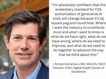
How will generative AI software be evaluated by the FDA in the future? What’s next for point-of-care ultrasound? What steps have been successful in facilitating adoption of AI applications across enterprise systems? Leaders in radiology and health care will be discussing these topics and more at the upcoming HLTH USA conference.
Current Perspectives on Integrating AI into Radiology
In a recent interview with Diagnostic Imaging, Joseph Cavallo, MD, MBA, offered insights on post-deployment monitoring with AI, the potential for improved workflow efficiency and the ongoing challenge of balancing essential clinical skills and AI literacy in the training of radiology residents and fellows.