The Value of MRI After Traumatic Brain Injury
Several lesion patterns were significantly associated with poor long-term neurocognitive and psychiatric outcomes.
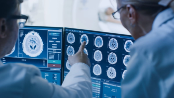
MRI following traumatic brain injury yields important prognostic information identifying patients who may have poor long-term neurocognitive and psychiatric outcomes, according to a study published in the
Researchers from Saudi Arabia and Egypt sough to identify the radiological and clinical factors associated with the functional capacity one year after the traumatic brain injury (TBI).
A total of 251 patients with severe head trauma were enrolled in the study. All underwent brain MRI within one month after injury. Demographic, clinical, and radiological data were collected during hospitalization. Neurocognitive and psychiatric evaluation were done one year thereafter.
The results showed that diffuse axonal injury was more frequent in the study patients. Psychiatric disorders, cognitive impairment, and poor functional outcome were more common in patients with DAI-especially those with cerebral hemisphere and brain stem lesion, and mixed lesions, the authors wrote. Duration of post traumatic amnesia, lost consciousness, and hospital stay, as well as the volume of diffuse axonal injury were associated with poor neurocognitive outcome. The duration of post traumatic amnesia and DAI may be considered independent factors that could predict the neurocognitive outcome.
The researchers concluded that by administering an MRI following traumatic brain injury, physicians will determine important prognostic information, with several lesion patterns significantly associated with poor long-term neurocognitive and psychiatric outcomes.
Newsletter
Stay at the forefront of radiology with the Diagnostic Imaging newsletter, delivering the latest news, clinical insights, and imaging advancements for today’s radiologists.


