|Articles|February 1, 2009

Diagnostic Imaging

  • Diagnostic Imaging Vol 31 No 2
  • Volume 31
  • Issue 2

3D neuro imaging techniquerealizes Orwellian vision

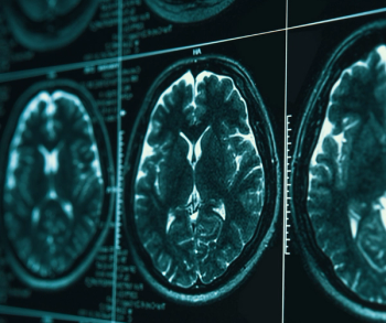
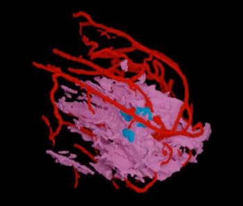
University of California, San Diego scientists are developing a new imaging modality that will study the body/brain dynamics of humans engaged in normal activity.

University of California, San Diego scientists are developing a new imaging modality that will study the body/brain dynamics of humans engaged in normal activity.

The Swartz Center for Computational Neuroscience at UCSD is creating the concurrent brain and body imaging modality MoBI (Mobile Brain/Body Imaging) under a four-year $3.4 million research grant from the U.S. Navy Office of Naval Research. According to principal investigator Scott Makeig, Ph.D., the insight provided by fMRI could be used by MoBI to “read” motivations behind actions by performing cognitive monitoring. If successful, the technique will allow functional studies outside the confined laboratory setting, such as screening the brain activity of workers in stressful situations.


Related to this article

Foundation Model-Based Chest X-Ray AI Software Gets FDA Nod
Reportedly one of the first FDA-cleared chest X-ray AI software platforms derived from a foundation model, the Chest Xray software facilitates detection and localization of lung nodules, pneumothorax, mediastinal abnormalities and other thoracic abnormalities.
Does kV Still Matter in Photon-Counting CT?
Emerging research demonstrates the increasing stability of attenuation on clinically relevant VMIs with photon-counting CT and a redefined role of kV that may have implications for longitudinal quantitative CT.