Breast CT enters realm of diagnosis and therapy
Developers of CT scanners have been trying to adapt their technology to breast imaging for decades. They have drawn a step closer, thanks to the efforts of researchers at the University of California, Davis. Their efforts may lead to the ability to not only visualize but treat breast cancer.
Developers of CT scanners have been trying to adapt their technology to breast imaging for decades. They have drawn a step closer, thanks to the efforts of researchers at the University of California, Davis. Their efforts may lead to the ability to not only visualize but treat breast cancer.
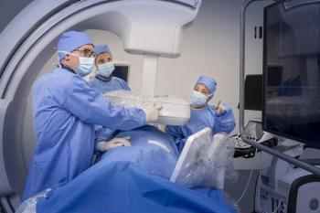
The UCD research team has spent the last ¬five years developing a breast CT scanner. Their prototypic detector rotates around the breast suspended through an opening in the patient table, collecting data that are reconstructed into 3D images. The total radiation dose given the 200 women scanned with the device so far has been no more than they would have received if they had undergone conventional mammography.
Earlier this year the researchers, led by John M. Boone, UCD professor and vice chair of radiology, reported progress in leveraging the CT cone beam to go a step further: not only diagnosing breast cancer and targeting treatment, but possibly even killing the malignant cells that remain after interventional therapy.
CT scans of the breast hold the potential to guide interventional procedures, such as robotic biopsy, radiofrequency ablation, and cryoablation, Boone said. More intriguing, however, is the possibility that the CT beams might be used to destroy cancers. The California researchers have measured dose distribution and performed simulations to show the feasibility of such a system, presenting their data about such therapeutic and targeting applications last summer at the American Association of Physicists in Medicine meeting in Anaheim, CA.
Computer simulations and physical phantom-based experiments determined the dose distributions for radiation therapy that might be achieved using the breast CT system. Several beam energies, from 120 kVp to 480 kVp, were simulated. In addition to homogeneous dose distributions for treating the whole breast, focused therapeutic approaches using collimators may be possible, according to preliminary results. The pendent position of the breast, which occurs when the patient is prone, may also create a more reproducible approach for fractionated radiotherapy of the breast.
Newsletter
Stay at the forefront of radiology with the Diagnostic Imaging newsletter, delivering the latest news, clinical insights, and imaging advancements for today’s radiologists.


