
|Articles|January 1, 2007
Exam distinguishes benign from malignant lesions
Author(s)C. P. Kaiser
Breast ultrasound elastography allows radiologists to accurately distinguish benign from malignant breast lesions. The technique correctly identified both cancerous and benign lesions in nearly 125 cases studied.
Advertisement
Breast ultrasound elastography allows radiologists to accurately distinguish benign from malignant breast lesions. The technique correctly identified both cancerous and benign lesions in nearly 125 cases studied.
"In our work, elasticity imaging has been found to have high specificity," said Dr. Richard G. Barr, a professor of radiology at Northeastern Ohio Universities College of Medicine in Youngstown. "If our results can be reproduced in a large multicenter trial, this technique could significantly reduce the number of breast biopsies required."
Barr evaluated 166 lesions with a real-time freehand elasticity imaging technique. Images were obtained using modified Siemens Elegra or Antares ultrasound systems; Barr is a consultant for Siemens. Lesions in which the elasticity image was smaller than the fundamental image were characterized as benign. Lesions in which the elasticity image was larger were characterized as malignant.
Ultrasound-guided biopsies were performed on 123 lesions. Biopsy showed that elasticity imaging correctly identified all 17 malignant lesions and 105 of 106 benign lesions, for a sensitivity of 100% and a specificity of 99%.
While mammography remains the gold standard exam for breast cancer screening, screening with MRI or ultrasound may be more effective for high-risk patients or women with dense breast tissue. MRI and ultrasound depict more breast lesions than mammography but have low specificity, resulting in a high number of invasive biopsies. About 80% of breast lesions biopsied are found to be benign, according to the American Cancer Society.
"Our ability to find lesions in the breast has increased significantly over the last 10 years but at the expense of an increased number of biopsies," Barr said. "This technique could significantly reduce the number of biopsies and increase the confidence of women that a detected lesion is truly benign."
He anticipates that elasticity imaging will also help in cancer detection but did not evaluate that capability for this study. The research will be expanded to an international multicenter trial beginning in January 2007.
Advertisement
Related Content
Advertisement


Advertisement
Advertisement
Trending on Diagnostic Imaging
1
New Study Shows Substantial Decline in CT Radiation Dosing for Adults Over the Past Decade
2
FDA Grants De Novo Authorization for AI-Based Coronary Inflammation Quantification from CCTA Exams
3
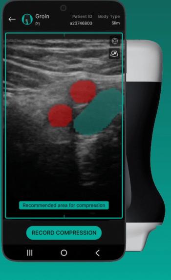
FDA Clears Ultrasound Guidance Software on Vascular Imaging for DVT Evaluation
4
Can AI-Enabled Intratumoral CT Threshold Segmentation Help Predict Visceral Pleural Invasion in Lung Cancer Patients?
5