|Articles|November 11, 2002
CAD and PACS take their place in the new millennium
Acceptance of computer-aided diagnosis has been growing steadily for more than 30 years, with FDA approval either achieved or pending for CAD devices related to chest radiographs, chest CT, and mammography. "We anticipate that the rapid advance of
Advertisement
Acceptance of computer-aided diagnosis has been growing steadily for more than 30 years, with FDA approval either achieved or pending for CAD devices related to chest radiographs, chest CT, and mammography.
"We anticipate that the rapid advance of these technologies will continue, and that application will broaden to cover much of medical imaging," said Dr. Bradley Erickson, a radiologist at the Mayo Clinic in Rochester.
Acceptance and integration of CAD technology with the electronic radiology practice is a current challenge, Erickson said in a paper (J Digit Imaging 2002;Sept 26:epub) discussing CAD at the start of the new century.
"If CAD is to become an integral tool for image interpretation, it is crucial that it become integrated into PACS workflow," he said. "These challenges will be overcome, and we expect that computer-aided diagnosis will be routinely applied to medical images."
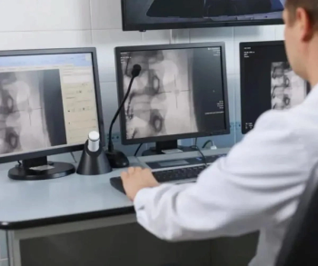
According to Erickson, PACS is important for CAD and vice versa. PACS implies digital acquisition and review. Digitizing films is labor-intensive and probably loses some information, both of which result in a significant penalty for CAD. While CAD has some demonstrated value, that value may be outweighed by losses in efficiency if images are not digital and there is a separate system for reviewing the CAD results.
CAD can also help drive PACS, as some areas - mammography, for example - have high data demands and perhaps provide less value for image sharing than other imaging methods.
But if CAD drives digital image acquisition, PACS will have additional justification and payback.
CAD is often viewed as a threat to radiologists, who fear that it will replace them, but Erickson believes the opposite is true.
First, in light of the predicted continuing radiologist shortages, getting some help may be a good thing, he said.
"But more important, the tasks that computers are good at -well-defined things like 'this is a normal chest X-ray' or 'this brain tumor is now 15.2 cc and has enlarged 0.6 cc since the last exam' - are things that humans typically do not enjoy," he said.
On the other hand, the problem-solving required for difficult cases is often what physicians enjoy most and what computers are least able to do.
"Therefore, I believe CAD will help filter the uninteresting cases that we don't need to put effort into and give us time to really focus on the challenging cases," he said.
Advertisement
Related Content
Advertisement


Advertisement
Advertisement
Trending on Diagnostic Imaging
1
Can AI Enhance Non-Contrast and Contrast-Enhanced CT Detection of Pancreatic Cancer?
2
Emerging PET Radiotracer May Enhance Detection of Small Metastases in Patients with Advanced Melanoma
3
Can Photon-Counting CT Provide Timely Clarity After Mechanical Thrombectomy?
4
Can Biomechanical CT Have an Impact in Opportunistic Screening for Osteoporosis?
5