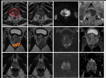
There were no statistical differences between bpMRI and mpMRI with respect to specificity with different EPE features such as psuedocapsule (PC) bulging (91 percent vs. 92 percent), PC irregularity (88 percent vs. 89 percent) and PC breach (99 percent for both), according to the study authors. The researchers also noted low sensitivity for these morphological criteria for both bpMRI and mpMRI.1
“ … bpMRI and mpMRI demonstrated comparable diagnostic performance for the assessment of EPE features. While both protocols were highly specific, neither was sufficiently sensitive for reliable detection of microscopic or subtle EPE. Within the limitations of this selected cohort, these findings suggest that omission of DCE does not substantially impair MRI assessment of EPE and support bpMRI as an alternative to mpMRI for local staging in comparable clinical settings,” noted lead study author Leon M. Bischoff, MD, who is affiliated with the Department of Diagnostic and Interventional Radiology at University Hospital Bonn and the University of Bonn in Bonn, Germany, and colleagues.
The study authors also noted no significant differences in sensitivity nor specificity with bpMRI and mpMRI in detecting neurovascular bundle invasion and seminal vesicle invasion.1
Does mpMRI Offer an Advantage with Detection of csPCa?
However, the researchers noted that the use of mpMRI demonstrated consistently higher sensitivity for clinically significant prostate cancer (csPCa) among the four reviewing radiologists in comparison to bpMRI (an average 79.75 percent for mpMRI vs. 76.25 percent for bpMRI).1
“These findings emphasize the diagnostic value of incorporating dynamic DCE sequences alongside T2-weighted and diffusion-weighted imaging, particularly for enhancing lesion conspicuity and reducing uncertainty in equivocal cases. However, large prospective data suggest this added value of DCE sequences translates only into marginal differences in clinically significant cancer detection, with bpMRI shown to be non-inferior to mpMRI,” added Bischoff and colleagues.1,2
Three Key Takeaways
• bpMRI performs comparably to mpMRI for EPE staging. No statistically significant difference emerged between the two protocols for overall EPE detection (AUC ~79.6 percent vs. 79.3 percent), or for specific EPE features like pseudocapsule bulging, irregularity, and breach. These findings suggest that the omission of dynamic contrast-enhanced (DCE) MRI sequences doesn't meaningfully compromise local staging accuracy.
• Both protocols share the same sensitivity limitation. Regardless of which sequence set is used, neither bpMRI nor mpMRI reliably detects microscopic or subtle EPE. Accordingly, a negative EPE read on either protocol shouldn't be over-relied upon to rule out extraprostatic disease.
• mpMRI retains an edge specifically for csPCa lesion detection, not staging. DCE sequences modestly boosted sensitivity for clinically significant prostate cancer detection (~79.75 percent vs. 76.25 percent) across readers so the choice between protocols may hinge more on lesion detection needs (and reader experience with equivocal cases) than on EPE assessment itself.
(Editor’s note: For related content, see “Meta-Analysis Shows Prognostic Impact of Prostate MRI Prior to Radical Prostatectomy,” “Can an Integrated Approach with PSMA PET/CT and Prostate MRI Enhance Detection of Extraprostatic Extension?” and “Can AI Enhance PET/MRI Assessment for Extraprostatic Tumor Extension in Patients with PCa?”)
Beyond the inherent limitations of a single-center retrospective study, the authors acknowledged a high prevalence of clinically significant prostate cancer and MRI-visible lesions within the 90-patient cohort. The researchers also noted a lack of clarity for how DCE MRI-only signs affect PI-RADS v2.1 categorization of EPE.
References
- Bischoff LM, Schmidt CJ, Endler C, et al. Comparison of biparametric and multiparametric prostate MRI to assess extraprostatic tumor extension. Eur J Radiol. 2026 July 23. In press. Available at: https://doi.org/10.1016/j.ejrad.2026.113110
2. Twilt JJ, Saha A, Bosma JS, et al. Evaluating biparametric versus multiparametric magnetic resonance imaging for diagnosing clinically significant prostate cancer: an international, paired, noninferiority, confirmatory observer study. Eur Urol. 2025;87(2):240-250.