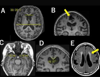
“During the follow-up period, participants with mild PD displayed progressive cortical thinning in the left hemisphere (mainly in the temporal and parietal lobes), with no change in GM volumes. This atrophy reflects the distribution of misfolded α-synuclein and subsequent progressive neuronal loss. … Notably, our GM results highlight left hemispheric lateralization, consistent with literature showing left hemisphere susceptibility in PD and in neurodegenerative diseases in general,” wrote lead study author Silvia Basaia, M.D., who is affiliated with the Neuroimaging Research Unit within the Division of Neuroscience at the IRCCS San Raffaele Scientific Institute in Milan, Italy, and colleagues.
In contrast to prior studies, the study authors noted they were able to evaluate changes to cortical thickness and volume in the brain over a three-year period and noted a clear correlation between DE indexes of structural connectivity and functional connectivity.
“In other words, changes in physical connections between different regions of the brain appear to be related to changes in how these regions functionally communicate,” explained Basaia and colleagues.
Three Key Takeaways
1. Predictive value of MRI-based connectome mapping for gray matter atrophy in Parkinson's disease. Brain MRI, specifically through connectome mapping based off diffusion tensor imaging and resting-state functional MRI, can help predict the progression of gray matter (GM) atrophy in patients with mild Parkinson's disease (PD).
2. Correlation of disease exposure Indexes with GM atrophy. Structural and functional disease exposure (DE) indexes correlate with the progression of GM atrophy over time, particularly in the right caudate nucleus and regions of the frontal, temporal, and parietal lobes.
3. Left hemisphere vulnerability in the brain. The study notes progressive cortical thinning in the left hemisphere of the brain in PD patients, which is consistent with previous literature on the susceptibility of the left hemisphere in neurodegenerative diseases.
In addition to the right caudate nucleus, the researchers noted the prognostic accuracy of GM atrophy progression with some of the temporal, parietal, and frontal brain regions.
“Early temporal lobe atrophy and subsequent frontal and parietal lobe degeneration may serve as biomarkers for the development of multidomain cognitive impairment and progression to PD with mild cognitive impairment. Furthermore, cortical thinning in frontal, temporal, and parietal regions and the hippocampus and thalamus is associated with conversion to more severe cognitive impairment in PD,” emphasized Basaia and colleagues.
While noting that “gray matter atrophy is an indirect measure of neurodegeneration, Kei Yamada, M.D., Ph.D., in an accompanying editorial, said the study authors clearly demonstrated a staged progression with Parkinson’s disease.
“(The researchers) were successful in showing that there is indeed a correlation between the functional and structural organization of the brain and the pattern of cortical atrophy,” wrote Dr. Yamada, who is affiliated with the Department of Radiology at Kyoto Prefectural University of Medicine in Kyoto, Japan.
(Editor’s note: For related content, see “Essential Keys to MRI Safety in the Age of Advanced Diagnostics,” “Video Interview: Is There an Increased Incidence of Neurodegenerative Diseases in Patients with COVID-19?” and “Emerging AI Software for Brain MRI Gets FDA Nod.”)
In regard to study limitations, the authors acknowledged using a 1.5T magnetic field and noted challenges with false-positive rates when employing a false discovery rate to assess changes in gray matter.