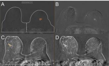
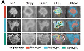
“Active surveillance is a rising paradigm for DCIS management under which participants with DCIS at low risk of upstaging and future invasive events do not receive treatment and are instead mammographically monitored,” wrote Kalina P. Slavkova, Ph.D., who is affiliated with the Department of Radiology at Columbia University Medical Center in New York City, and colleagues.
“For successful implementation of active surveillance, it is imperative that selection criteria have a high sensitivity to minimize harm while reducing overtreatment. Reliable models for risk stratification would help standardize selection criteria and ultimately optimize DCIS management.”
Three Key Takeaways
1. Enhanced risk stratification. The integration of breast MRI-based radiomic features with clinical and qualitative MRI factors improves the prediction of disease upstaging in women with DCIS, offering a more reliable tool for risk assessment.
2. Improved specificity. The combined model incorporating radiomic features demonstrated a 25 percent higher specificity (53 percent vs. 28 percent) compared to using only clinical and qualitative MRI features, potentially reducing unnecessary treatments.
3. Support for active surveillance. Reliable risk stratification models could help refine criteria for active surveillance, reducing overtreatment while ensuring high sensitivity in identifying low-risk DCIS cases suitable for non-interventional management.
Emphasizing that the radiomic features in this study were derived from standard-of-care imaging, the researchers suggested that continued research into biomarkers could further enhance the identification of viable candidates for active surveillance.
“ … Additional biomarkers that can help improve prediction of low-risk cases, such as novel pathologic features, should be explored to further decrease risks of choosing active surveillance and expand the number of participants eligible for de-escalation,” posited Slavkova and colleagues.
(Editor’s note: For related content, see “Breast MRI and Dense Breasts: A Closer Look at Early Findings from a New Prospective Trial,” “Mammography-Based AI Abnormality Scoring May Improve Prediction of Invasive Upgrade of DCIS” and “Can DWI MRI Offer a Viable Non-Contrast Alternative for Breast Cancer Assessment?”)
In regard to study limitations, the authors acknowledged the use of manual lesion localization and segmentation. The researchers noted variability with the timing of initial post-contrast images and conceded that later post-contrast phases are more advantageous for observing the commonly persistent kinetics of DCIS in comparison to the study’s emphasis on early post-contrast phases. The study authors also pointed out the absence of qualitative MRI and histopathologic data for a proportion of the cohort.