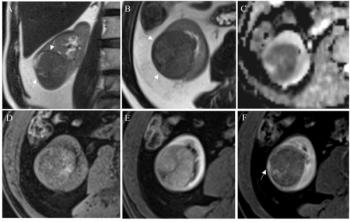
In time-independent testing for 36 patients, the researchers noted a 37.5 percent higher sensitivity (79.2 percent) with EOB-MRI’s modified diagnosis algorithm in contrast to ECA-MRI (41.7 percent).
“EOB-MRI with a modified algorithm exhibited superior diagnostic performance for sub-centimeter recurrent HCC compared with ECA-MRI, providing a more sensitive and reliable imaging strategy for early recurrence identification,” noted lead study author Yuyao Xiao, MD, who is affiliated with the Shanghai Institute of Medical Imaging and the Department of Radiology at Zhongshan Hospital in Shanghai, China, and colleagues.
Three Key Takeaways
• EOB-MRI significantly improves sensitivity for sub-centimeter recurrent HCC detection. A modified diagnostic algorithm for gadoxetic acid-enhanced MRI achieved markedly higher sensitivity (~79–81 percent) compared to extracellular contrast MRI (~42 percent), while maintaining high specificity.
• Diagnostic tradeoff favors EOB-MRI for early recurrence detection. While the modified diagnosis algorithm for ECA-MRI demonstrated very high specificity (~98 percent), its low sensitivity limits utility for early detection. The modified diagnosis algorithm for EOB-MRI offers a more balanced and clinically useful performance profile for identifying small recurrent lesions.
Incorporating transitional phase washout enhances detection beyond current LI-RADS guidance. Expanding washout assessment to include the transitional phase — along with T2WI mild or moderate hyperintensity and diffusion restriction — substantially boosts sensitivity without compromising specificity, suggesting a potential refinement to existing imaging criteria.
Noting that current LI-RADS guidelines only recommend non-peripheral washout assessment on the portal venous phase (PVP) for EOB-MRI, the study authors emphasized that evaluating washout in the transitional phase (TP) with EOB-MRI can yield dividends with sub-centimeter HCC detection.
“… Previous studies suggest that including TP in the washout assessment can improve sensitivity without compromising specificity, even in sub-centimeter HCC. In our study, EOB-MRI-based modified algorithm—the combination of T2WI mild-moderate hyperintensity, non-peripheral washout on PVP or TP and restricted diffusion—showed significantly improved sensitivity compared with the typical vascular pattern … while also maintaining high specificity,” added Xiao and colleagues.
(Editor’s note: For related content, see “Emerging MRI Scoring System May Help Predict Recurrent and Metastatic Hepatocellular Carcinoma,” “Could Contrast-Enhanced Ultrasound Offer Equivalency to MRI for Detecting Small HCC Nodules?” and “Multicenter Study Affirms Value of Updated AASLD Criteria for Surveillance for Hepatocellular Carcinoma.”)
Beyond the inherent limitations of a single-center retrospective trial, the authors cautioned that the cohort — drawn from a Chinese tertiary center with a majority of patients having HBV-related HCC — may thwart extrapolation of the study findings to broader populations.