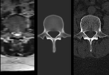
“The proposed open-source, easy-to-use model allows for automatic, robust segmentation of MRI scans, independent of MRI sequence, and achieved good performance across 80 structures … ,” wrote lead study author Tugba Akinci D’Antonoli, M.D., who is affiliated with the Clinic of Radiology and Nuclear Medicine at University Hospital Basel in Basel, Switzerland, and colleagues.
In a separate ablation analysis, the researchers found that the combination of MRI and CT training with the TotalSegmentator MRI-3 segmentation model offered a higher Dice score than another model trained on MRI only (86.2 percent vs. 84.5 percent).
Three Key Takeaways
1. High segmentation accuracy. The TotalSegmentator MRI model demonstrated strong performance, achieving an 83.9 percent Dice score across 80 anatomical structures, outperforming MRSegmentator and AMOS segmentation models in specific testing cohorts.
2. Enhanced performance with CT training data. Training the model with both MRI and CT data led to improved segmentation accuracy (86.2 percent Dice score vs. 84.5 percent for MRI-only training), suggesting that CT data enhances MRI segmentation robustness.
3. Clinical integration and research potential. The study authors maintained that the open-source segmentation model can be seamlessly integrated into clinical workflows for real-time assistance in radiology and has potential applications in research, such as studying organ size variations based on age and disease.
“This finding indicates that incorporating CT images in the training dataset enhances the model’s performance for MRI segmentation and effectively serves as a form of data augmentation, a common method in deep learning to improve model robustness,” posited D’Antonoli and colleagues.
(Editor’s note: For related content, see “AI Segmentation, Intraprostatic Tumor Volume and Metastases: What a New mpMRI Study Reveals,” “FDA Clears New AI-Powered Spine MRI Software” and “AI Software for Brain MRI Gets Expanded FDA Clearance for Multiple Sclerosis Assessment.”)
The study authors noted the potential of the TotalSegmentator MRI model for assessing organ size based on factors such as age and disease state and emphasized its current availability (http://totalsegmentator.com ) as a viable alternative for MRI segmentation.
“Our model can be easily integrated into existing clinical workflows and can operate in real time to assist radiologists during diagnostic processes,” noted D’Antonoli and colleagues. “It can also be used in various research projects, for example, analyzing age-dependent changes in the volume of different abdominal structures.”