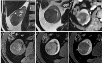
“DWI can provide information on pathological and physiological changes earlier than conventional MRI by reflecting the diffusion characteristics of water molecules in tissues and can be used for early diagnosis and evaluation of efficacy at the cellular level,” wrote Xinyu Li, M.D., who is affiliated with the First Affiliated Hospital College of Clinical Medicine of the Henan University of Science and Technology in Henan, China, and colleagues.
For DCE-MRI, the researchers noted a 73 percent AUC, 78 percent sensitivity and 65 percent specificity for gauging patient response to concurrent chemoradiotherapy. In assessing patient response to neoadjuvant therapy, the study authors found that DCE-MRI had a 70 percent AUC, an 84 percent sensitivity rate and a 61 percent specificity rate.
Three Key Takeaways
1. DWI MRI's efficacy. Diffusion-weighted MRI (DWI MRI) demonstrated high accuracy in predicting response to concurrent chemoradiotherapy and neoadjuvant therapy in esophageal cancer, with an AUC of 88 percent, sensitivity of 80 to 82 percent, and specificity of 81 percent.
2. DCE-MRI as a supplementary tool. Dynamic contrast-enhanced MRI (DCE-MRI) was less accurate than DWI MRI with an AUC ranging from 70 percent for neoadjuvant therapy response to 73 percent for concurrent chemoradiotherapy response. However, the meta-analysis authors noted the potential of DCE-MRI for assessing tissue changes and neovascularization in esophageal cancer.
3. Predictive value of MRI. Pre- and post-treatment DWI MRI and DCE-MRI assessments can provide insights into the likelihood of treatment response, with DWI showing an 87 percent probability of non-response when no pathological changes are seen.
“DCE can rapidly and continuously scan and shows a dynamic continuous enhancement through digital reconstruction technology, thereby showing the continuous change of tumor tissue. According to the flow and distribution of contrast agents in tumor tissue, the changes in tumor tissue morphology and function can be analyzed, and neovascularization can often be seen in esophageal cancer tissue,” noted Li and colleagues.
When pre- and post-treatment DWI MRI didn’t reveal any pathological response to concurrent chemoradiotherapy, the study authors noted an 87 percent probability of no treatment response. The researchers noted a 70 percent probability of no response to concurrent chemoradiotherapy without evidence of pathological response in pre- and post-treatment DCE MRI assessments.
(Editor’s note: For related content, see “MRI Features Help Detect Neoadjuvant Therapy Response in Esophageal SCC,” “Image IQ Quiz: Headaches in a 55-Year-Old Patient with Esophageal Cancer” and “Image IQ Quiz: Patient with History of Excessive Drinking Vomits Blood.”)
In regard to study limitations, the authors conceded that differences in chemotherapy regimens, contrast agent injection and the timing of image acquisition during treatment can affect MRI assessments of treatment for esophageal cancer.