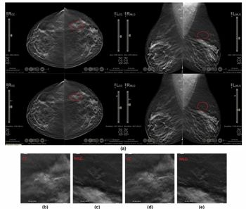
Is There a Question of Surgeon Confidence in CEM?
The researchers found no differences between those women who had CEM and those who did not with respect to mastectomy rates (17 percent vs. 18 percent) and reoperation rates (7.9 percent vs. 10 percent).
However, they suggested that surgeon preferences may have been a key factor. Noting that surgeons at one of the reviewing facilities adhered to breast-conserving treatment in most cases, the study authors said the surgeons relied more on digital mammography (DM) and digital breast tomosynthesis (DBT) than CEM to determine malignancy extent.
“This approach contributed to the absence of evidence of a difference in reoperation rates between the CEM and control groups,” maintained Gardfjell and colleagues.
Three Key Takeaways
• Preoperative CEM changed surgical management in a meaningful minority of patients. Of 202 women who had preoperative CEM, 17 had their treatment changed, and histopathology confirmed 16 of those changes. Most changes were escalations from partial mastectomy to mastectomy (eight cases). These changes occurred regardless of menopausal status, breast density, or lesion type, so CEM’s added value is not limited to dense breasts.
• CEM estimated tumor extent more accurately than the other modalities. CEM agreed more closely with histopathology (bias of 3.5 mm) than DM/DBT (4.8 mm) or ultrasound (10.7 mm). This held overall and in almost all subgroups. That makes CEM a strong option for preoperative sizing and surgical planning.
• The benefit did not show up in overall outcomes, which may reflect surgeons’ trust in CEM. Mastectomy rates (17 percent vs. 18 percent) and reoperation rates (7.9 percent vs. 10 percent) were similar with and without CEM. The authors partly attribute this to surgeons at one of the study sites who relied on DM/DBT rather than CEM findings for malignancy extent and favored breast-conserving surgery. CEM’s impact may depend on multidisciplinary confidence with the modality.
What the Evidence Shows About CEM and Agreement with Malignancy Extent on Histopathology Results
Yet there was closer agreement between CEM and the extent of malignancy on histopathology results (bias, 3.5 mm) in contrast to that for DM and DBT (4.8 mm) and breast ultrasound (10.7 mm), according to the study authors.
“Estimation of malignant extent was also superior with CEM compared with digital mammography, digital breast tomosynthesis, and US, overall and in almost all subgroups. With increased clinical confidence in CEM, results suggest that preoperative CEM may help optimize treatment for many patients with breast cancer,” added Gardfjell and colleagues.
(Editor’s note: For related content, see “The Reading Room Podcast: Current Perspectives, Challenges and Possibilities with AI in Breast Imaging, Part 2,” “Video: Stamatia Destounis, MD, FACR, Discusses Key Changes for the Updated BI-RADS System” and “Emerging Research in Mammography, Breast MRI, Ultrasound and AI: A Summer Roundup of Key Breast Imaging Studies.”)
In regard to study limitations, the authors acknowledged possible bias with the unblinded study design. They also conceded a lack of availability for CEM-guided biopsy, differences in surgical approach that may have impacted treatment decisions and insufficiently powered analysis for detecting subgroup differences with pre-op CEM.