Register Now: Image-Based Risk: The Next Frontier in Breast Cancer Screening
Blog|Articles|May 1, 2026

Why Photon-Counting CT is the New Standard for Non-Invasive Stent Imaging

The benefits of photon-counting CT via ultra-high spatial resolution, reduced blooming artifacts and high temporal resolution allow characterization with confidence in coronary stent assessment.

For years, coronary stent evaluation by computed tomography angiography (CTA) was criticized due to the amount of blooming or metal artifact and often dismissed with “Just send the patient to invasive angiography.

That paradigm belongs to the past. Photon-counting CT (PCCT) is changing non-invasive stent imaging from a limitation into a true diagnostic tool.

Why does conventional CT struggle?

With standard energy-integrating detector CT (EID-CT), metallic struts create significant blooming, partial volume effects obscure the intra-stent lumen, small stents become nearly unreadable and motion artifacts worsen proximal and ostial evaluation.

Subsequently, many stents are labeled “non-diagnostic” and patients are sent to unnecessary invasive angiography.

How does PCCT change everything?

• Ultra-highspatial resolution provides sharper visualization of stent struts and enables clear assessment of the true intra-stent lumen.
• With reduced blooming artifacts, metal appears closer to reality and there is less artificial lumen narrowing.
• High temporal resolution allows better imaging of proximal, ostial, and fast-moving segments along with reduced motion blur around the stent.

• High iodine concentration contrast (400 mg I/mL) offers strong intraluminal enhancement, better contrast between lumen and metal, and spectral capability.
• There is additional confidence in separating iodine signal from stent artifact.

The clinical shift goes from “Can we read this stent?” to “We can characterize it with confidence.”
Restenosis, stent patency, aneurysm repair, complex proximal LAD interventions and left main and bifurcation stents all become more reliably assessable.
From invasive default to non-invasive confidence and convenience, from metallic blur to diagnostic clarity … that’s why PCCT is becoming the new standard for coronary stent imaging.

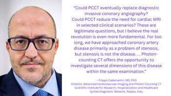
Dr. Cademartiri is the director of advanced cardiovascular imaging and photon-counting CT at the Scientific Institute for Research, Hospitalization, and Healthcare Synlab Diagnostic Network in Naples, Italy. He is also a consultant in advanced cardiovascular imaging at CDI/Centro Diagnostico Italiano in Milan, Italy.

(Editor’s note: This blog is adapted with permission from Dr. Cademartiri’s original LinkedIn post at:

https://www.linkedin.com/feed/update/urn:li:activity:7455133379529875456/ .)


Related to this article

Does kV Still Matter in Photon-Counting CT?
Emerging research demonstrates the increasing stability of attenuation on clinically relevant VMIs with photon-counting CT and a redefined role of kV that may have implications for longitudinal quantitative CT.
CT Radiation Dosing: Where Things Stand, Part 2
In the second of a two-part interview, Kalpana Kanal, PhD, and Mahadevappa Mahesh, MS, PhD, FAPPM, FACR, discussed future directions for research on CT radiation dosing, including the potential of indication-specific diagnostic reference levels (DRLs).
Emerging Research Insights and New Advances in Neuroradiology
Catch up on key neuroradiology news and research from the past month, including recently published studies involving white matter hyperintensity findings on brain MRI, MRI-enhanced amyloid PET and the newly FDA-approved PET agent floretyrosine F18 for glioma imaging.