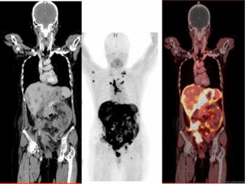
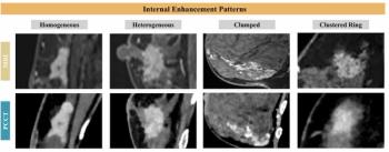
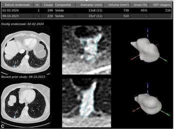
New research demonstrates significant differences between fluorodeoxyglucose-positron emission tomography (FDG-PET) and computed tomography (CT) in the assessment of response for patients being treated for metastatic breast cancer.
For the retrospective study, recently published in the European Journal of Radiology, researchers compared CT scans (assessed with the RECIST 1.1 criteria) and FDG-PET imaging (evaluated with the PET-based PERCIST criteria) in 51 patients who received systemic treatment for metastatic breast cancer. Out of a total of 375 scans acquired for these patients between 2017 and 2025, there were 151 time-matched pairs of CT and FDG-PET scans, according to the study.
Is There an Overreliance on the CT-Based RECIST 1.1 Criteria?
The study authors found that 65 percent of the paired CT scans (98/151) failed to provide measurable target lesions, according to the RECIST 1.1 criteria. In contrast, the researchers noted that all paired FDG-PET images (151/151) offered measurable target lesions.
“ …More than one-third of CT examinations in our cohort could not be categorized according to RECIST 1.1, primarily due to bone-only disease or lymph nodes below established size thresholds,” pointed out lead study author Vilma Landfors Andersson, MD, who is affiliated with the Faculty of Medicine at Lund University in Lund, Sweden, and colleagues.
When comparing CT and FDG-PET imaging that did reveal measurable target lesions, the researchers noted a 79 percent agreement on lesion location. However, the study authors said the concordance on target-lesion identification plummeted to 28 percent when assessing all 151 pairs of CT and FDG-PET scans.
Three Key Takeaways
• RECIST 1.1 (CT) frequently fails to capture measurable disease in metastatic breast cancer. Sixty-five percent of paired CT scans lacked measurable target lesions — mainly due to bone-only disease or sub-threshold lymph nodes — versus 0 percent of FDG-PET/PERCIST scans. This suggests that CT alone may be inadequate for staging or trial eligibility in a substantial share of patients.
• CT and FDG-PET often disagree on which lesions to track. Researchers noted that concordance on target-lesion identification dropped to just 28 percent across all paired scans (79 percent when only scans with measurable CT lesions were compared). This raises concerns about consistency in response assessment depending on modality choice.
• FDG-PET detects progression earlier than CT. FDG-PET flagged progressive disease a median of 50 days sooner and identified earlier progression in 61 percent of cases with CT never outperforming PET in timing. This supports a potential role for FDG-PET in more timely treatment evaluation and clinical trial patient selection.
Does FDG-PET Identify Disease Progression Earlier than CT in Cases of Metastatic Breast Cancer?
The researchers also determined that FDG-PET identified progressive metastatic disease at a median of 50 days earlier than CT. Overall, in the time-matched scan pairs, the study authors found that FDG-PET detected disease progression earlier than CT in 61 percent of cases whereas CT did not detect progression earlier than FDG-PET in any of the cases.
“ … These findings provide real-world insight into how imaging modality influences response assessment and disease classification in (metastatic breast cancer). They highlight limitations of CT-based criteria, including implications for clinical trial eligibility, and suggest a potential role for FDG-PET in improving treatment evaluation and patient selection for clinical trials,” added Andersson and colleagues.
(Editor’s note: For related content, see “Researchers Show Higher Breast Cancer Upstaging with 18F-FAPI PET/CT,” “Can Radioligand Therapy Have an Impact for Women with Breast Cancer?” and “Emerging Insights on the Use of FES PET for Women with Lobular Breast Cancer.”)
Final Notes
Beyond the inherent limitations of a non-randomized retrospective study, the authors acknowledged the small cohort size and potentially inflated discordance between negative CT scan and PET imaging with cases involving predominantly osseous and/or nodal disease having a higher likelihood of being referred for FDG-PET.