The study authors also found that employing an O-RADS US 4 score as a minimum threshold for detection of malignancy yielded a 94 percent sensitivity rate and a 98 percent negative predictive value (NPV).
While noting the high reliability of the O-RADS US risk scoring system for excluding malignant diagnoses, the researchers cautioned that the O-RADS US system has drawbacks in specificity (64 percent) and positive predictive value (PVV) (38 percent).
“This decreased specificity and PPV limit the use of O-RADS US version 2022 for predicting the presence of malignancy in this patient population,” noted Shen and colleagues.
Three Key Takeaways
1. Potential to reduce unnecessary surgery. Use of the O-RADS US 2022 risk stratification system could have prevented surgery in nearly half of the studied patients with ovarian cystic lesions, offering them follow-up imaging or conservative management instead.
2. High sensitivity and NPV for malignancy. Employing an O-RADS US 4 score as the minimum threshold for malignancy detection yielded a 94 percent sensitivity and a 98 percent negative predictive value (NPV), demonstrating its reliability in ruling out malignant ovarian lesions.
3. Limitations in specificity. While O-RADS US is reliable in excluding malignancy, its lower specificity (64 percent) and positive predictive value (38 percent) limit its accuracy in predicting malignancy. MRI may be useful for further characterization of uniocular cysts scored as O-RADS US 2 and lesions scored as O-RADS US 3 or higher..
In an accompanying editorial, Laure S. Fournier, M.D., Ph.D., praised the originality of the study in assessing the practical application and role of the O-RADS US system within the management of patients with ovarian and adnexal lesions.
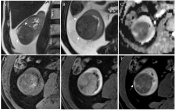
In regard to the lower specificity of O-RADS US, Fournier said subsequent magnetic resonance imaging (MRI) can enhance characterization of uniocular cysts scored as O-RADS US 2 as well as other lesions with O-RADS US 3 or higher risk scores.
“Moreover, MRI allows for identification of certain histologic types, which helps the surgeon know what to expect, prepare the procedure, and inform the patient on the possible findings,” added Dr. Fournier, a professor in the Department of Radiology at the Georges Pompidou European Hospital in Paris, France.
(Editor’s note: For related content, see “Key Challenges with the O-RADS Ultrasound Classification System,” “Is MRI More Effective than Ultrasound for Diagnosing Adnexal Lesions?” and “MRI or Ultrasound for Evaluating Pelvic Endometriosis?: Seven Takeaways from a New Literature Review.”)
In regard to study limitations, the authors noted the exclusion of patients with acute presentations at the time of ultrasound examination but still acknowledged possible patient selection bias with the likelihood of a higher PPV in a cohort of patients undergoing surgery. The researchers also noted that ultrasound scans were obtained by technologists who lacked training in gynecologic imaging and reviewed by radiologists in academic settings in contrast to non-academic practices where physicians with gynecologic imaging experience perform and interpret the scans.