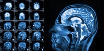
However, the presence of THC on baseline MRI was associated with a nearly threefold higher risk for cognitive decline within five to seven years. The researchers noted the elevated risk was evident in the entire cohort as well as those deemed to be cognitively normal in baseline testing.
“These findings suggest that careful assessment of THC may aid early risk stratification. Future studies incorporating longitudinal imaging will be needed to clarify the mechanisms underlying THC and to establish its role in early risk stratification for cognitive decline,” noted lead study author Kana Saito, MD, who is affiliated with the Department of Radiology at the Hirosaki University Graduate School of Medicine in Hirosaki, Japan, and colleagues.
Three Key Takeaways
• Tightness of the high-convexity sulci (THC) on brain MRI may be a useful early warning sign. In this prospective study of 2,133 community-dwelling older adults (median age 69), THC at baseline was linked to a nearly threefold higher risk of cognitive decline over five to seven years. The association held in participants who were cognitively normal at baseline, which suggests the finding could help with risk stratification before symptoms appear.
• THC combined with Sylvian fissure enlargement may be more informative than traditional DESH criteria. People who had both THC and enlarged Sylvian fissures (ESF) were 83 percent more likely to have long-term cognitive decline. The authors suggest this pairing may be a more clinically meaningful marker than the Evans index or complete DESH. Complete DESH was rare in the cohort (1.7 percent), while incomplete DESH was common (46.3 percent). This supports assessing individual DESH features rather than only the full triad.
• The findings are promising but not yet ready for routine use. Cognitive outcomes were based on clinical diagnosis of MCI or dementia rather than standardized neuropsychological testing. The cohort was also limited to older Japanese adults from a single region, which may restrict generalizability. The authors call for longitudinal imaging studies to clarify how THC develops and to confirm its role in early risk stratification.
The study authors also noted an 83 percent increase in the likelihood of long-term cognitive decline for participants in the total cohort who had THC and ESF on brain MRI.
“ … These findings indicate that ESF with THC may serve as a more clinically meaningful marker than (the Evans index) or complete DESH for identifying older individuals at risk for cognitive decline,” added Saito and colleagues.
(Editor’s note: For related content, see “Emerging Research Insights and New Advances in Neuroradiology,” “Can Brain MRI Surveillance Alone Be Advantageous in Monitoring Cognition for Patients with SCLC?” and “What New CT and MRI Research May Reveal About Bone Loss, Diabetes and Cognitive Decline.”)
In regard to study limitations, the authors noted the use of clinical diagnosis of MCI or dementia for cognitive assessments as opposed to standard neuropsychologic battery testing, and a cohort entirely comprised of older Japanese patients from a single demographic region.